. 2011 Jan 26;31(4):1375-85.
doi: 10.1523/JNEUROSCI.2791-10.2011.

Activation of TREK currents by the neuroprotective agent riluzole in mouse sympathetic neurons

Affiliations

Activation of TREK currents by the neuroprotective agent riluzole in mouse sympathetic neurons

Alba Cadaveira-Mosquera et al. J Neurosci. .

Abstract

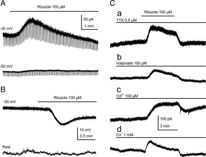
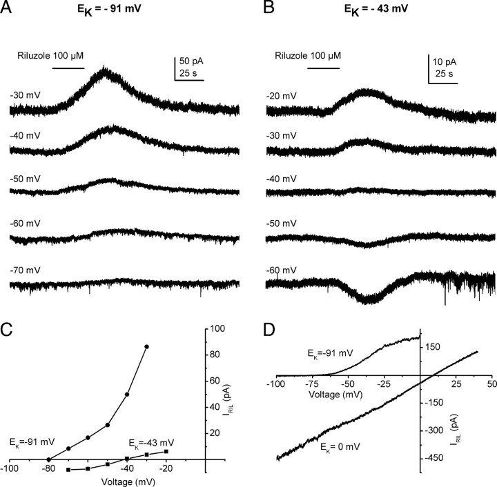
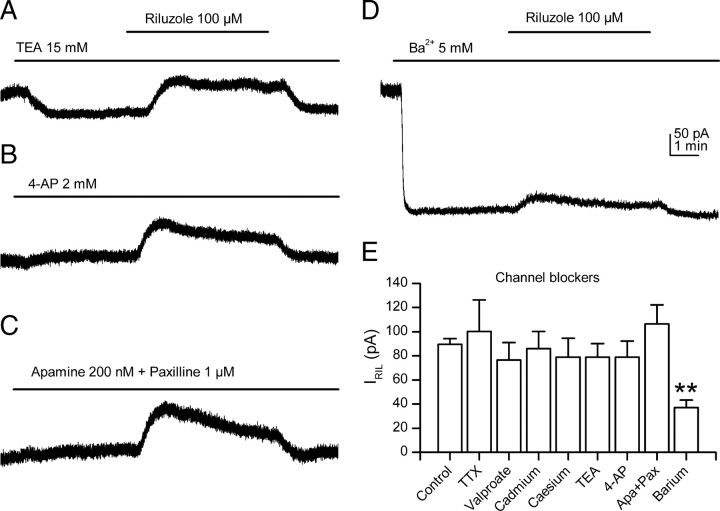
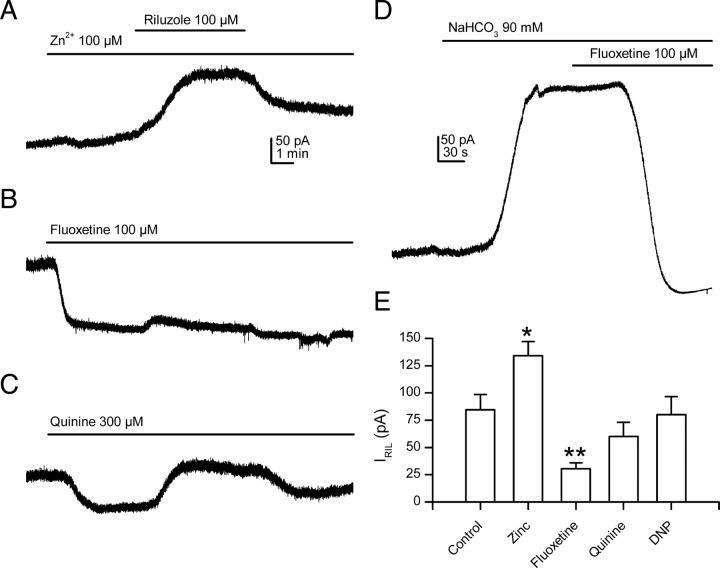
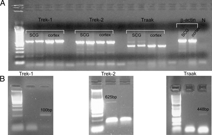
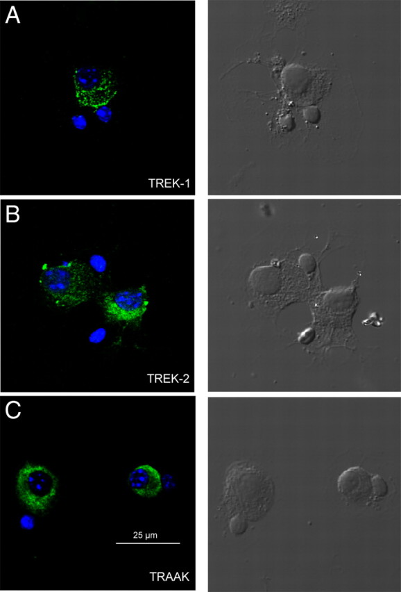
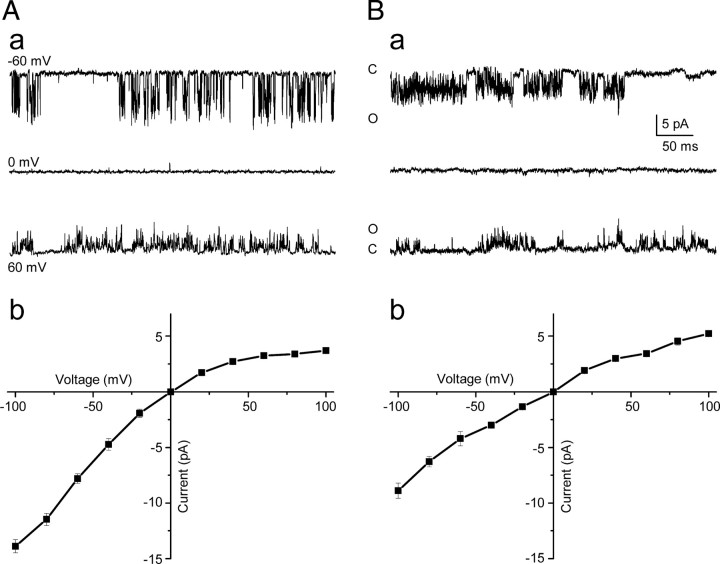
Background K2P channels play a key role in stabilizing the resting membrane potential, thereby modulating cell excitability in the central and peripheral somatic nervous system. Whole-cell experiments revealed a riluzole-activated current (I(RIL)), transported by potassium, in mouse superior cervical ganglion (mSCG) neurons. The activation of this current by riluzole, linoleic acid, membrane stretch, and internal acidification, its open rectification and insensitivity to most classic potassium channel blockers, indicated that I(RIL) flows through channels of the TREK [two-pore domain weak inwardly rectifying K channel (TWIK)-related K channel] subfamily. Whole-ganglia and single-cell reverse transcription-PCR demonstrated the presence of TREK-1, TREK-2, and TRAAK (TWIK-related arachidonic acid-activated K(+) channel) mRNA, and the expression of these three proteins was confirmed by immunocytochemistry in mSCG neurons. I(RIL) was enhanced by zinc, inhibited by barium and fluoxetine, but unaffected by quinine and ruthenium red, strongly suggesting that it was carried through TREK-1/2 channels. Consistently, a channel with properties identical with the heterologously expressed TREK-2 was recorded in most (75%) cell-attached patches. These results provide the first evidence for the expression of K2P channels in the mammalian autonomic nervous system, and they extend the impact of these channels to the entire nervous system.


Figures

Figure 1.
Figure 1.
Riluzole activates an outward current in mSCG neurons. A, Application of riluzole induced a clear transient outward current when the membrane was held at −30 mV (top trace). The amplitude of IRIL was strongly reduced at more negative membrane potentials (bottom trace). Short-duration hyperpolarizing voltage pulses were continuously applied to follow changes in conductance. B, In neurons manually current clamped at −30 mV, riluzole application induced a clear membrane hyperpolarization (top trace), an effect that was strongly reduced or absent at resting membrane potentials (bottom trace). C, The peak amplitude of the riluzole-activated current was unaffected by blockers of voltage-dependent sodium channels (a), persistent sodium channels (b), voltage-activated calcium channels (c), and hyperpolarization-activated cationic channels (d).
Figure 2.
Figure 2.
IRIL is a potassium current with a reversal potential that follows the Nernst equation. A, In physiological external potassium concentrations (3 mm), short applications of riluzole evoked increasing outward currents at potentials more positive than −80 mV. In most cells tested, no inward currents could be obtained at more negative voltages. B, Inward currents were obtained at potentials below to −40 mV when the extracellular potassium concentration was elevated to 20 mm. C, Current–voltage relationships for the riluzole-induced currents shown in A and B. Note that the marked outward rectification in 3 mm potassium (dots) was strongly reduced in 20 mm potassium (squares). In both cases, the reversal potential of IRIL was close to the Nernst equilibrium potential for potassium. D, Current–voltage relationships for riluzole-induced currents obtained in response to negatively progressing voltage ramps. The current obtained in the control was subtracted from that obtained in the presence of riluzole (100 μm) (close to the peak). Note that the strong outward rectification obtained in standard solutions (EK = −91 mV) disappeared when symmetrical concentrations of potassium were used (EK = 0 mV). Recordings were acquired from two different cells.
Figure 3.
Figure 3.
IRIL is transported through K2P channels. A–C, The riluzole-induced current was not affected by the presence of classic potassium channel blockers TEA, 4-AP, apamine, and paxilline, indicating that IRIL is not transported through voltage- or calcium-activated potassium currents. D, Barium clearly and significantly reduced the outward current evoked by riluzole. The scale bars in this figure apply to all the panels. E, Bar diagram summarizing the main results obtained using ion channel blockers. This figure represents the mean ± SEM for the peak current induced by 100 μm riluzole at a holding potential of −30 mV. **p < 0.01 versus control.
Figure 4.
Figure 4.
TREK but not TRAAK channels provide the main contribution to IRIL. A, Application of zinc to mSCG neurons induced a slowly developing outward current and significantly increased the riluzole-activated current. B, Fluoxetine induced an inward current at −30 mV and strongly reduced IRIL. C, Quinine also provoked an inward current, but the effect on the riluzole-activated current was not statistically significant. D, Internal acidification by extracellular bicarbonate resulted in the activation of an outward current that was completely blocked by fluoxetine. E, Peak amplitude of riluzole activated currents in the presence of modulators of TREK channels. *p < 0.05 and **p < 0.01 versus control. All the experiments in this figure were carried in the blockers extracellular solution, and the membrane was held at −30 mV. The scale bars in A apply for B and C.
Figure 5.
Figure 5.
mSCG neurons express mRNAs encoding all the members of the TREK subfamily. A, Expression of mRNAs for the TREK channels in the whole mSCG and cerebral cortex as assessed by RT-PCR using specific primers for each K2P channel. DNA fragments were cloned and sequenced for confirmation. The expected band sizes were 678 bp (TREK-1, lanes 2, 3, 4, 5), 625 bp (TREK-2, lanes 7, 8, 9, 10), 448 bp (TRAAK, lanes 12, 13, 14, 15), and 655 bp (β-actin, lanes 17, 18). The first lane shows the 100 bp ladder (Takara). Negative controls are shown in lanes 6, 11, 16, and 19. B, Expression of TREK-1 (left), TREK-2 (middle), and TRAAK (right) mRNA in individual mSCG cells assessed by single-cell RT-PCR.
Figure 6.
Figure 6.
mSCG neurons express the three protein subunits of the TREK subfamily. A–C, Specific FITC-conjugated antibodies against TREK-1 (A), TREK-2 (B), and TRAAK (C) stained cultured mSCG neurons (green, left panels). DAPI-stained nuclei (blue) not surrounded by green stain belong to satellite cells that often accompanied the neurons in the culture. Satellite cells were never positive for FITC, and they can be observed under Nomarski optics in the right panels.
Figure 7.
Figure 7.
Large-conductance TREK-2 potassium channels were recorded in most cell-attached patches. Aa, TREK-2 cell-attached single channel currents recorded at −60, 0, and +60 mV in symmetrical 150 KCl solutions. Ab, Mean (±SEM) current–voltage relationships for 12 TREK-2 channels showing distinctive inward rectification. Ba, TREK-like single channel activity at −60, 0, and +60 mV in symmetrical solutions. Note the flickering and bursting behavior characteristic of TREK channels. Bb, Mean (±SEM) current–voltage relationships for three TREK-like channels showing weaker inward rectification. C, Closed; O, open.

Similar articles

Cited by

References

    1. Altschul SF, Gish W, Miller W, Myers EW, Lipman DJ. Basic local alignment search tool. J Mol Biol. 1990;215:403–410. - PubMed
    1. Bang H, Kim Y, Kim D. TREK-2, a new member of the mechanosensitive tandem-pore K+ channel family. J Biol Chem. 2000;275:17412–17419. - PubMed
    1. Brown DA, Constanti A. Intracellular observations on the effects of muscarinic agonists on rat sympathetic neurones. Br J Pharmacol. 1980;70:593–608. - PMC - PubMed
    1. Brown DA, Adams PR, Constanti A. Voltage-sensitive K-currents in sympathetic neurons and their modulation by neurotransmitters. J Auton Nerv Syst. 1982;6:23–35. - PubMed
    1. Buckler KJ, Vaughan-Jones RD. Effects of mitochondrial uncouplers on intracellular calcium, pH and membrane potential in rat carotid body type I cells. J Physiol. 1998;513:819–833. - PMC - PubMed

Publication types

MeSH terms

LinkOut - more resources