. 2011 Jan 26;31(4):1419-26.
doi: 10.1523/JNEUROSCI.4259-10.2011.

Crystal structure of the amyloid-β p3 fragment provides a model for oligomer formation in Alzheimer's disease

Affiliations

Crystal structure of the amyloid-β p3 fragment provides a model for oligomer formation in Alzheimer's disease

Victor A Streltsov et al. J Neurosci. .

Abstract

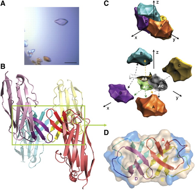
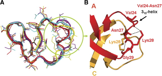
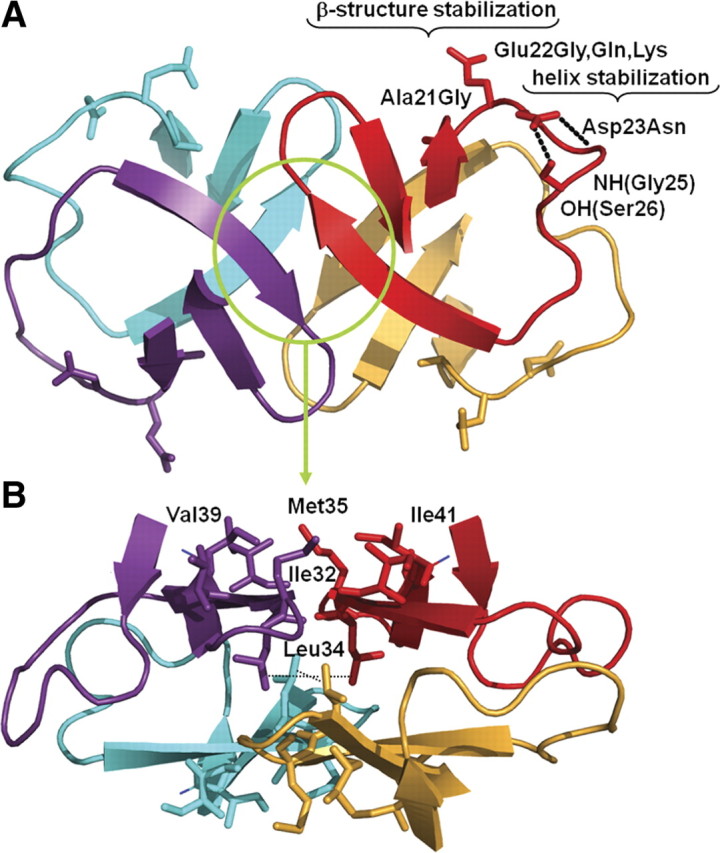
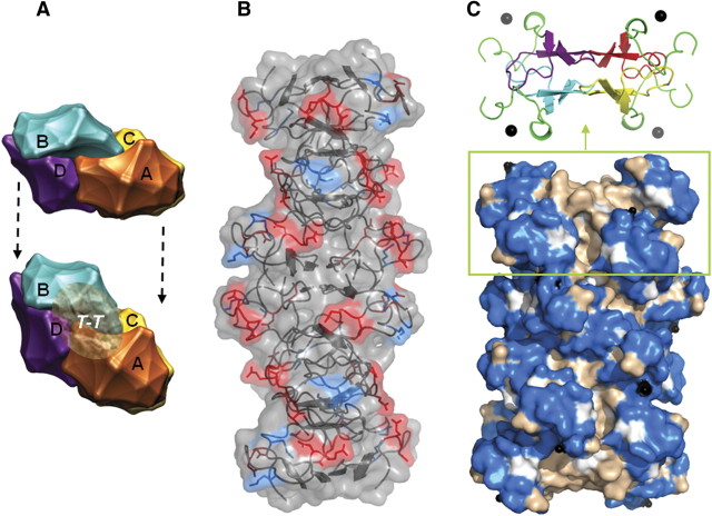
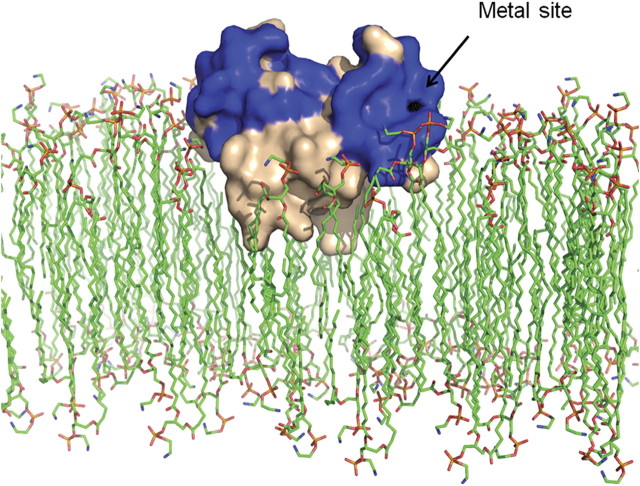
Alzheimer's disease is a progressive neurodegenerative disorder associated with the presence of amyloid-β (Aβ) peptide fibrillar plaques in the brain. However, current evidence suggests that soluble nonfibrillar Aβ oligomers may be the major drivers of Aβ-mediated synaptic dysfunction. Structural information on these Aβ species has been very limited because of their noncrystalline and unstable nature. Here, we describe a crystal structure of amylogenic residues 18-41 of the Aβ peptide (equivalent to the p3 α/γ-secretase fragment of amyloid precursor protein) presented within the CDR3 loop region of a shark Ig new antigen receptor (IgNAR) single variable domain antibody. The predominant oligomeric species is a tightly associated Aβ dimer, with paired dimers forming a tetramer in the crystal caged within four IgNAR domains, preventing uncontrolled amyloid formation. Our structure correlates with independently observed features of small nonfibrillar Aβ oligomers and reveals conserved elements consistent with residues and motifs predicted as critical in Aβ folding and oligomerization, thus potentially providing a model system for nonfibrillar oligomer formation in Alzheimer's disease.


Figures

Figure 1.
Figure 1.
Crystal structure of the Aβ-IgNAR-G1 tetramer. A, Aβ-IgNAR-G1 crystallized under a wide variety of conditions, in this instance 200 mm TMAO (trimethylamine N-oxide), 100 mm Tris, pH 8.5, 20% PEG MME (monomethyl ether) 2000. Scale bar, 100 μm. B, Diagram representation of four IgNAR domains (color washed) caging Aβ (in rectangular box). C, Aβ tetramer interaction surfaces. D, The four Aβ chains (A–D) interact across six β-sheet structures. Semitransparent solubility surface is shown as hydrophilic (marine blue) and hydrophobic (light pink) areas.
Figure 2.
Figure 2.
Aβ peptide chains structure. A, Aβ peptide loops overlay, illustrating chain divergence between residues Val24–Asn27 (circled). The Lys28 side chain is oriented differently in chains A/B compared with chains C/D. B, Conformations of Lys28 for chains A and C illustrating hydrogen bond and electrostatic interactions.
Figure 3.
Figure 3.
Correlation with familial AD and in vitro mutation data. A, Residues Ala21, Glu22, and Asp23 are exposed within a β-turn. B, Residue Leu34 occupies a pivotal position within both the dimer and tetramer interfaces. Hydrophobic residues Ile32, Met35, Val39, and Ile41, important in amyloid formation in this model, are shown at the top of the structure.
Figure 4.
Figure 4.
Models of amyloid multimeric oligomer formation. A, Assembly of the Aβ oligomer from two tetramers with the T-T interface. B, Aβ oligomer model based on six Aβ18–41 tetramers. Overlaid surfaces highlight negatively charged (red) and positively charged (blue) residues. C, As for A and B, incorporating Aβ1–17 metal-binding regions (solubility surfaces: marine blue, hydrophilic; light pink, hydrophobic). The black spheres represent metals (Zn, Cu, etc.). The N-terminal Aβ1–17 is in green in the inset diagram.
Figure 5.
Figure 5.
Model of potential interactions of Aβ18–41 dimer with membrane lipid bilayers. The hydrophobic dimer–dimer interface of the Aβ18–41 tetramer is intercalated into the membrane surface through nonelectrostatic interactions, whereas hydrophilic aspects (blue) with metal-binding sites (black) are on the membrane surface.

References

    1. Barghorn S, Nimmrich V, Striebinger A, Krantz C, Keller P, Janson B, Bahr M, Schmidt M, Bitner RS, Harlan J, Barlow E, Ebert U, Hillen H. Globular amyloid β-peptide oligomer—a homogenous and stable neuropathological protein in Alzheimer's disease. J Neurochem. 2005;95:834–847. - PubMed
    1. Barnham KJ, Cappai R, Beyreuther K, Masters CL, Hill AF. Delineating common molecular mechanisms in Alzheimer's and prion diseases. Trends Biochem Sci. 2006;31:465–472. - PubMed
    1. Baumketner A, Krone MG, Shea JE. Role of the familial Dutch mutation E22Q in the folding and aggregation of the 15–28 fragment of the Alzheimer amyloid-β protein. Proc Natl Acad Sci U S A. 2008;105:6027–6032. - PMC - PubMed
    1. Betts V, Leissring MA, Dolios G, Wang R, Selkoe DJ, Walsh DM. Aggregation and catabolism of disease-associated intra-Aβ mutations: reduced proteolysis of AβA21G by neprilysin. Neurobiol Dis. 2008;31:442–450. - PMC - PubMed
    1. Brünger AT. Free R value: a novel statistical quantity for assessing the accuracy of crystal structures. Nature. 1992;355:472–475. - PubMed

MeSH terms

Substances