. 2011 Mar;25(3):394-408.
doi: 10.1210/me.2010-0458. Epub 2011 Jan 27.

Ligand-dependent degradation of SRC-1 is pivotal for progesterone receptor transcriptional activity

Affiliations

Ligand-dependent degradation of SRC-1 is pivotal for progesterone receptor transcriptional activity

Larbi Amazit et al. Mol Endocrinol. 2011 Mar.

Abstract

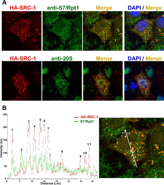
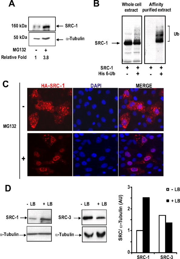
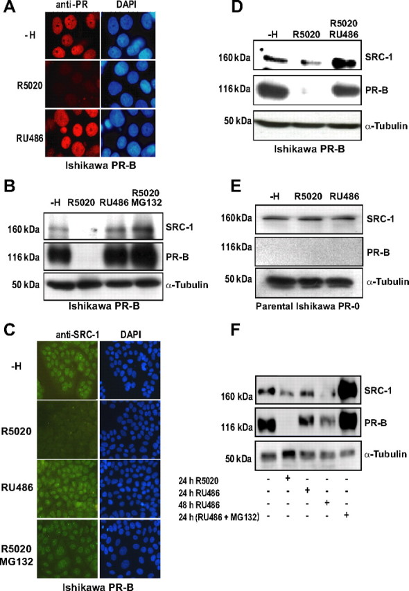
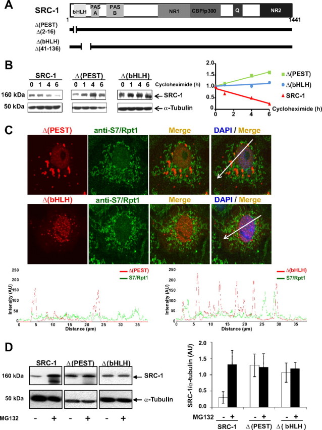
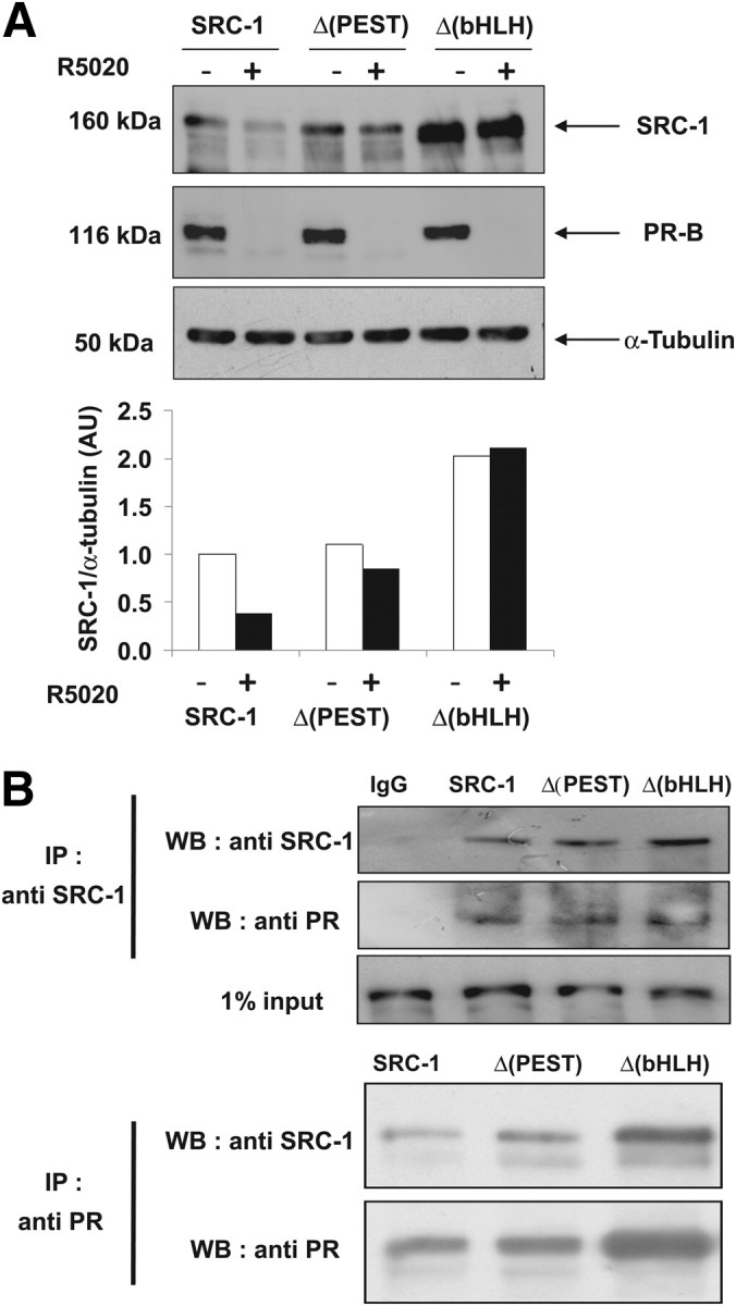
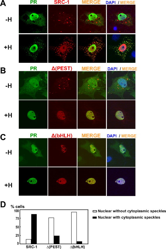
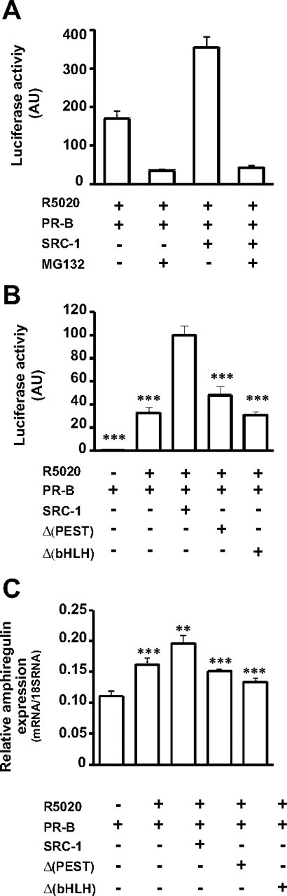
The progesterone receptor (PR), a ligand-activated transcription factor, recruits the primary coactivator steroid receptor coactivator-1 (SRC-1) gene promoters. It is known that PR transcriptional activity is paradoxically coupled to its ligand-dependent down-regulation. However, despite its importance in PR function, the regulation of SRC-1 expression level during hormonal exposure is poorly understood. Here we report that SRC-1 expression level (but not other p160 family members) is down-regulated by the agonist ligand R5020 in a PR-dependent manner. In contrast, the antagonist RU486 fails to induce down-regulation of the coactivator and impairs PR agonist-dependent degradation of SRC-1. We show that SRC-1 proteolysis is a proteasome- and ubiquitin-mediated process that, predominantly but not exclusively, occurs in the cytoplasmic compartment in which SRC-1 colocalizes with proteasome antigens as demonstrated by confocal imaging. Moreover, SRC-1 was stabilized in the presence of leptomycin B or several proteasomal inhibitors. Two degradation motifs, amino-acids 2-16 corresponding to a PEST motif and amino acids 41-136 located in the basic helix loop helix domain of the coactivator, were identified and shown to control the stability as well as the hormone-dependent down-regulation of the coactivator. SRC-1 degradation is of physiological importance because the two nondegradable mutants that still interacted with PR as demonstrated by coimmunoprecipitation failed to stimulate transcription of exogenous and endogenous target genes, suggesting that concomitant PR/SRC-1 ligand-dependent degradation is a necessary step for PR transactivation activity. Collectively our findings are consistent with the emerging role of proteasome-mediated proteolysis in the gene-regulating process and indicate that the ligand-dependent down-regulation of SRC-1 is critical for PR transcriptional activity.


Figures

Fig. 1.
Fig. 1.
Colocalization of SRC-1 with the 26S proteasome by confocal microscopy. A, Colocalization analysis between HA-SRC-1 and endogenous proteasome antigens S7/Rpt1 and 20S subunits. COS-7 cells were transiently transfected with the expression vector encoding HA-SRC-1. Cells were fixed after 40 h, immunolabeled with anti-HA and either anti-S7/Rpt1 or anti-20S antibodies, and then observed by confocal microscopy. B, Validation of colocalization by scan of intensity profiles of a representative cell [expressed as arbitrary units (AU)]. Fluorescence intensity was calculated and plotted by drawing a line through the middle of the cell image in a distance covering several cytosolic and nuclear foci. Green lines represent the intensity profile for the proteasome antigen S7/Rpt1 signal, and the red lines represent the intensity profile for SRC-1 signal. Indicated numbers refer to identified speckles: cytoplasmic (1 to 7) or nuclear (8 to 11). Note that although the fluorescence intensity from the two channels is different, the peaks of both signals are overlapping.
Fig. 2.
Fig. 2.
SRC-1 is proteolyzed by the 26S proteasome in a ubiquitin-dependent manner. A, COS-7 cells were transfected with the expression vector encoding SRC-1 and incubated in the absence or presence of MG132 (5 μm) during 15 h. Expression of SRC-1 was analyzed by Western blot using anti-SRC-1 and anti-α-tubulin antibodies. Band intensities corresponding to SRC-1 were quantified as described in Materials and Methods. B, CV-1 cells were transfected with the expression vector encoding HA-SRC-1 in the presence of the His6-tagged ubiquitin expression vector (His 6-Ub). Whole-cell extracts were analyzed by electrophoresis on 6.4% SDS-PAGE and immunoblotted with anti-HA monoclonal antibody. Alternatively, the same cotransfected CV-1 cells were lysed in buffer containing guanidium-HCl (Ni-NTA). The ubiquitin-modified proteins were purified using Ni-NTA agarose beads as described in Materials and Methods. Affinity-purified proteins were separated by electrophoresis, and His6-SRC-1 conjugates were detected by Western blot using the anti-HA monoclonal antibody. The ubiquitin conjugates of SRC-1 are indicated with brackets. C, COS-7 cells were transfected with the expression vector encoding HA-SRC-1. Twenty hours after transfection, cells were incubated during 24 h with MG132 (1 μm) or treated with vehicle (dimethylsulfoxide). Cells were then fixed and immunolabeled with anti-HA antibody. D, COS-7 cells were transfected with the expression vector encoding SRC-1 or SRC-3 and treated similarly than in C except that MG132 was replaced by LB treatment (20 ng/ml). Expression levels of SRC-1 and SRC-3 were analyzed by Western blot using anti-SRC-1 or anti-SRC-3 monoclonal antibodies as indicated. Band intensities representing the mean of at least two independent experiments were quantified as described in Materials and Methods.
Fig. 3.
Fig. 3.
Ligand- and PR-dependent SRC-1 proteolysis. A, Ishi PR-B cells, a cell line stably expressing PR-B, were cultured 24 h in the absence or in the presence of either the agonist R5020 (10 nm) or the antagonist RU486 (10 nm). Cells were then treated for immunocytochemistry with anti-PR antibody (Let 126) and observed by fluorescence microscopy. DAPI, 4′,6′-Diamino-2-phenylindole. B, Ishi PR-B cells were transfected with the SRC-1 encoding vector. After 48 h, cells were cultured 15 h as indicated, either in the absence of ligand (control vehicle, −H), in the presence of R5020 (10 nm) or RU486 (10 nm), or in the presence of both R5020 (10 nm) and MG132 (5 μm). Whole-cell extracts were analyzed by electrophoresis on 7.5% SDS-PAGE and immunoblotted with the indicated antibodies. C, Nontransfected Ishi PR-B cells were treated as in B. Cells were immunolabeled for endogenous SRC-1 using an anti-SRC-1 antibody. Note the agonist-ligand-dependent down-regulation of endogenous SRC-1. DAPI, 4′,6′-Diamino-2-phenylindole. D, Nontransfected Ishi PR-B cells were cultured 24 h in the absence of ligand (vehicle, −H) or in the presence of either the agonist R5020 (10 nm) alone or in combination with a 100× excess of the antagonist RU486 (1 μm). Whole-cell extracts were analyzed by electrophoresis on 7.5% SDS-PAGE and immunoblotted to detect endogenous SRC-1 and PR with the indicated antibodies. E, Ishi PR-0 cells (parental cell line, devoid of PR) were treated as in A. Cells were then analyzed by electrophoresis on 7.5% SDS-PAGE and immunoblotted with the indicated antibodies. F, Ishi PR-B cells were transfected with the SRC-1 encoding vector. After 24 h, cells were cultured in the absence of ligand (vehicle, −H), treated with R5020 (10 nm, 24 h) or RU486 (10 nm, 24 or 48 h) or RU486 (10 nm, 24 h) along with MG132 (1 μm). Whole-cell extracts were analyzed by electrophoresis on 7.5% SDS-PAGE and immunoblotted with the indicated antibodies.
Fig. 4.
Fig. 4.
The N-terminal region of SRC-1 targets the coactivator to degradation. A, Schematic representation of the wild-type coactivator SRC-1 (1441 amino acids in length) with boxes corresponding to major functional domains: PAS, Per-ARNT-Sim motif; NR1 and NR2, NR-interacting domains 1 and 2, CBP/p300 interacting domain; Q, glutamine-rich domain. SRC-1 deletion mutants Δ(PEST) and Δ(bHLH) are represented below with a thick line interrupted by a gap corresponding to the deleted amino acids. B, COS-7 cells were transfected as indicated with SRC-1, Δ(PEST), or Δ(bHLH) encoding vectors. Seventy-two hours after transfection, cells were treated with cycloheximide (100 μg/ml) during 1, 4, or 6 h. Whole-cell extracts were analyzed by electrophoresis on 7.5% SDS-PAGE and immunoblotted with the indicated antibodies. Band intensities (right panel) representing the mean of at least two independent experiments were quantified as described in Materials and Methods. C, Upper panel, Colocalization analysis of SRC-1 deletion mutants and S7/Rpt1. COS-7 cells were transiently transfected with Δ(PEST)- or Δ(bHLH)-encoding vectors. Cells were fixed after 40 h and immunolabeled with anti-HA and anti-Rpt1/S7 antibodies prior to analysis by confocal microscopy. Lower panel, Scan of intensity profiles expressed as arbitrary units (AU). Fluorescence intensity was calculated and plotted by drawing a line through the middle of the cell image in a distance covering several cytosolic and nuclear foci. Green lines represent the intensity profile for the proteasome antigen S7/Rpt1 signal, and the red lines represent the intensity profile for Δ(PEST) or Δ(bHLH) signals. Note the absence of significant peaks with overlapping signals. D, COS-7 cells were transfected with HA-SRC-1-, Δ(PEST)-, or Δ(bHLH)-encoding vectors. After 48 h, cells were incubated during 15 h with MG132 (5 μm) or vehicle. Whole-cell extracts were analyzed by electrophoresis on 7.5% SDS-PAGE and immunoblotted with the indicated antibodies. The band intensities (right panel) were quantified as described in Materials and Methods.
Fig. 5.
Fig. 5.
Ligand-dependent down-regulation of SRC-1 requires both degradation motifs of the coactivator. A, Ishi PR-B cells were transfected as indicated with HA-SRC-1-, Δ(PEST)-, or Δ(bHLH)-encoding vectors. After 48 h, cells were cultured in the absence of ligand (vehicle, −) or presence of the agonist R5020 (10 nm) during 24 h. The corresponding whole-cell extracts were analyzed by electrophoresis on 7.5% SDS-PAGE and immunoblotted with the indicated antibodies. The band intensities (lower panel) were quantified as described in Materials and Methods. B, HEK 293 cells were cotransfected with PR and the SRC-1-, Δ(PEST)-, or Δ(bHLH)-encoding vectors. Twenty four hours after transfection, cells were treated during 24 h with the agonist R5020 (10 nm). A coimmunoprecipitation assay was performed using the anti-SRC-1, the anti-PR, or the IgG1 control antibodies (IgG1). Purified proteins were separated on 7.5% SDS-PAGE. Coprecipitated complexes were identified with the indicated antibodies. IP, Immunoprecipitation; WB, Western blot.
Fig. 6.
Fig. 6.
Colocalization of PR and SRC-1 in cytoplasmic speckles. A, COS-7 cells were transiently transfected with the expression vector encoding HA-SRC-1 and PR. Twenty-four hours after transfection, cells were incubated or not for 8 h with R5020 in presence of cycloheximide (100 μg/ml) prior to fixation. Cells were immunolabeled with anti-PR (Let 126) and anti-HA antibodies. DAPI, 4′,6′-Diamino-2-phenylindole. B and C, cells were treated as in A, except that PR was transfected as indicated with Δ(PEST) and Δ(bHLH), respectively. D, Quantification of cells treated as described in A–C. Percent of cells treated with R5020 shows nuclear localization with or without cytoplasmic speckles. At least 100 cells were counted.
Fig. 7.
Fig. 7.
SRC-1 degradation is necessary for PR transcriptional activity. A, Ishi PR-0 cells were cotransfected as indicated with expression vectors encoding PR and SRC-1 together with the reporter gene PRE2-TATA-luc and the internal control pRS-β-gal. Cells were incubated with R5020 (10 nm) and treated or not with MG132 (500 nm) during 24 h. Luciferase activity was quantified and normalized by β-galactosidase activity. Data represent means ± sem of at least three independent determinations. B, COS-7 cells were cotransfected as indicated with HA-SRC1-, Δ(PEST)-, or Δ(bHLH)-encoding vectors together with expression vector encoding PR, the reporter gene PRE2-TATA-luc, and the internal control pRS-β-gal. Cells were treated during 24 h with R5020 (10 nm) or vehicle (control, −). Luciferase activity was quantified and normalized by β-galactosidase activity. Data represent means ± sem of four independent determinations performed in triplicate. C, Ishi PR-0 cells were cotransfected as indicated with HA-SRC-1-, Δ(PEST)-, or Δ(bHLH)-encoding vectors together with PR encoding vector and were treated with the agonist R5020 10 nm for 3 h. Total RNAs were extracted and relative expression of amphiregulin gene was quantified by quantitative RT-PCR. Results, normalized by the amplification of 18S RNA, are mean ± sem of three independent determinations. ***, Statistical significance, P < 0.001 vs. wild-type SRC-1 used as reference.

References

    1. Li X , O'Malley BW. 2003. Unfolding the action of progesterone receptors. J Biol Chem 278:39261–39264 - PubMed
    1. Lange CA. 2008. Challenges to defining a role for progesterone in breast cancer. Steroids 73:914–921 - PMC - PubMed
    1. Lonard DM , O'Malley BW. 2006. The expanding cosmos of nuclear receptor coactivators. Cell 125:411–414 - PubMed
    1. Xu J , Li Q. 2003. Review of the in vivo functions of the p160 steroid receptor coactivator family. Mol Endocrinol 17:1681–1692 - PubMed
    1. Chen D , Ma H , Hong H , Koh SS , Huang SM , Schurter BT , Aswad DW , Stallcup MR. 1999. Regulation of transcription by a protein methyltransferase. Science 284:2174–2177 - PubMed

Publication types

Substances