. 2011 Mar;25(3):503-15.
doi: 10.1210/me.2010-0340. Epub 2011 Jan 27.

ROS signaling by NOX4 drives fibroblast-to-myofibroblast differentiation in the diseased prostatic stroma

Affiliations

ROS signaling by NOX4 drives fibroblast-to-myofibroblast differentiation in the diseased prostatic stroma

Natalie Sampson et al. Mol Endocrinol. 2011 Mar.

Abstract

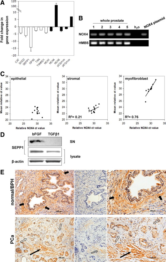
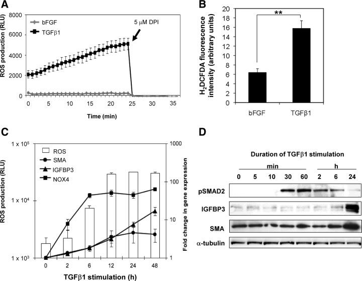
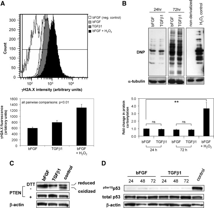
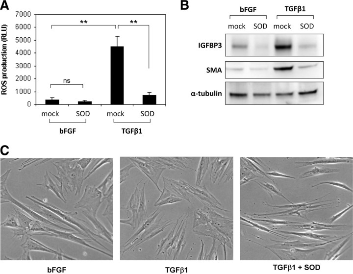
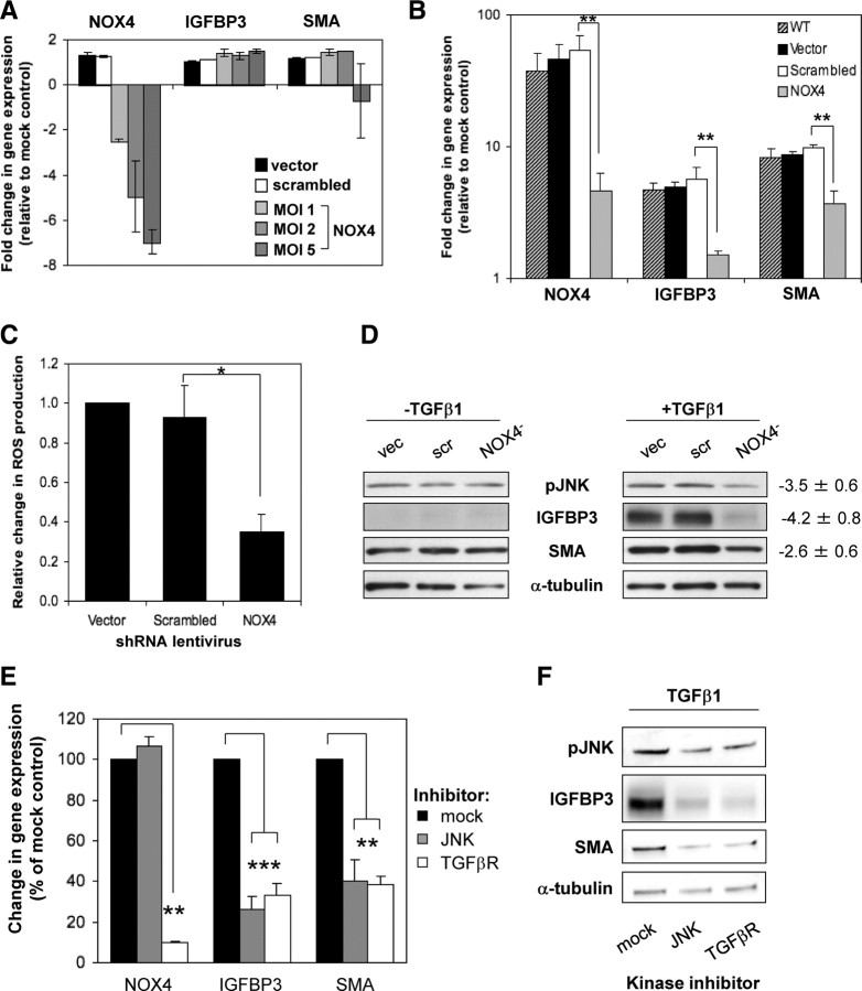
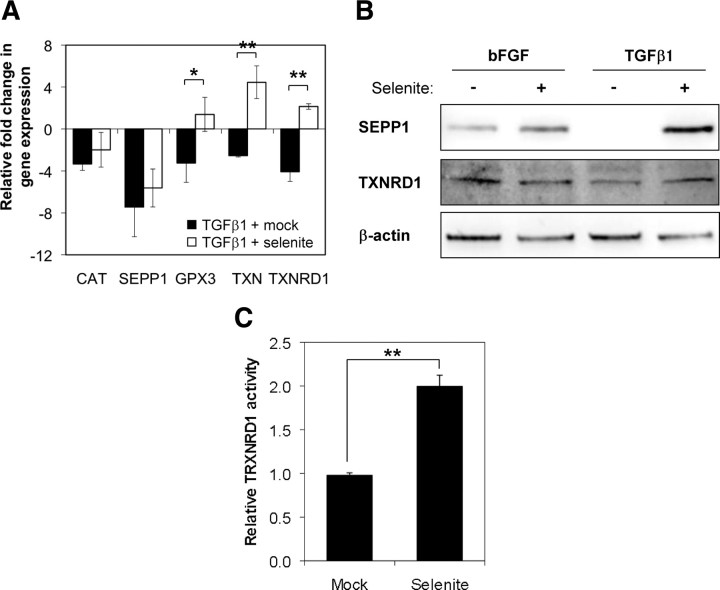
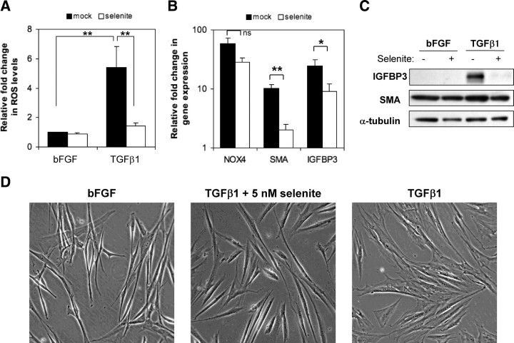
Stromal remodeling, in particular fibroblast-to-myofibroblast differentiation, is a hallmark of benign prostatic hyperplasia (BPH) and solid tumors, including prostate cancer (PCa). Increased local production of TGFβ1 is considered the inducing stimulus. Given that stromal remodeling actively promotes BPH/PCa development, there is considerable interest in developing stromal-targeted therapies. Microarray and quantitative PCR analysis of primary human prostatic stromal cells induced to undergo fibroblast-to-myofibroblast differentiation with TGFβ1 revealed up-regulation of the reactive oxygen species (ROS) producer reduced nicotinamide adenine dinucleotide phosphate oxidase 4 (NOX4) and down-regulation of the selenium-containing ROS-scavenging enzymes glutathione peroxidase 3, thioredoxin reductase 1 (TXNRD1), and the selenium transporter selenoprotein P plasma 1. Consistently, NOX4 expression correlated specifically with the myofibroblast phenotype in vivo, and loss of selenoprotein P plasma 1 was observed in tumor-associated stroma of human PCa biopsies. Using lentiviral NOX4 short hairpin RNA-mediated knockdown, pharmacological inhibitors, antioxidants, and selenium, we demonstrate that TGFβ1 induction of NOX4-derived ROS is required for TGFβ1-mediated phosphorylation of c-jun N-terminal kinase, which in turn is essential for subsequent downstream cytoskeletal remodeling. Significantly, selenium supplementation inhibited differentiation by increasing ROS-scavenging selenoenzyme biosynthesis because glutathione peroxidase 3 and TXNRD1 expression and TXNRD1 enzyme activity were restored. Consistently, selenium depleted ROS levels downstream of NOX4 induction. Collectively, this work demonstrates that dysregulated redox homeostasis driven by elevated NOX4-derived ROS signaling underlies fibroblast-to-myofibroblast differentiation in the diseased prostatic stroma. Further, these data indicate the potential clinical value of selenium and/or NOX4 inhibitors in preventing the functional pathogenic changes of stromal cells in BPH and PCa.


Figures

Fig. 1.
Fig. 1.
NOX4 and SEPP1 are associated with stromal remodeling in vivo. A, qPCR of ROS-scavenging (white bars) and ROS-producing (black bars) enzymes in PrSCs differentiated with 1 ng/ml TGFβ1 (48 h) relative to control cells incubated with 1 ng/ml bFGF (48 h) to maintain the fibroblast phenotype. Values represent mean fold change (±sem) of four independent experiments using different donors. B and C, NOX4 expression was evaluated in non-tumor-containing human prostate samples. B, RT-PCR of NOX4 (negative control using water as substrate; positive control using plasmid DNA containing full-length NOX4 cDNA). RT-PCR of HMBS is shown as loading control. C, qPCR of NOX4 in prostate specimens (n = 13) relative to the expression of epithelial, stroma, or myofibroblast markers as described in Materials and Methods. D, Western blotting of SEPP1 in lysates and supernatants (SN) of PrSCs treated with 1 ng/ml bFGF or TGFβ1 for 48 h. β-Actin is shown as loading control. A representative blot of three independent experiments is shown. E, SEPP1 immunohistochemistry (left) in normal/BPH and PCa biopsies (Gleason 7), enlarged images are shown (right), preincubation of anti-SEPP1 antibody with blocking peptide (center). Periglandular stromal cells (short black arrows), periglandular tumor stroma (open arrows), SMC bundles (long black arrow), weak immunostaining of SMCs due to incomplete blocking (gray arrow). Sections were counterstained with Mayer's hematoxylin. Tissue specimens were processed in parallel. Images are representative of four independent experiments with specimens from at least eight different donors.
Fig. 2.
Fig. 2.
Sustained ROS production precedes fibroblast-to-myofibroblast differentiation. A, ROS production was measured real-time in PrSCs 24 h after stimulation with TGFβ1 or bFGF as control via luminol-based chemiluminescence. Values represent mean of triplicate wells (±sem). A representative example of at least three experiments using independent donors is shown. B, ROS production was measured in PrSCs 48 h after stimulation with TGFβ1or bFGF via H2DCFDA staining and analyzed by fluorescence-activated cell sorting. Values represent mean fluorescence of triplicate samples using three different donors in independent experiments. Significance is indicated (**, P < 0.01). C, Time course assay of ROS production (left y-axis) and qPCR (right y-axis) of PrSCs stimulated for the indicated duration with TGFβ1. Mean values obtained from at least three experiments using independent donors are shown (±sem). D, Western blotting of lysates from PrSCs stimulated with TGFβ1 for the indicated duration with the antibody shown. Blots are representative of three independent experiments using different donors. RLU, Relative light units.
Fig. 3.
Fig. 3.
Elevated ROS production during differentiation do not induce major global DNA damage or protein oxidation. A, H2A.X phosphorylated at Ser139 was quantified via flow cytometry in PrSCs stimulated for 48 h with either bFGF or TGFβ1. Top panel, Histograms from a single experiment of γH2A.X staining intensity in PrSCs treated as indicated (negative control, omission of primary antibody in bFGF-treated samples). Note the increased (rightward) shift in staining intensity in H2O2 relative to bFGF and TGFβ1-treated samples. Lower panel, Mean values (±sem) of triplicate samples using different donors in three independent experiments. B (top panel), PrSCs were treated for the indicated duration with either bFGF or TGFβ1 before detection of total protein carbonyl levels via immunoblotting for anti-2,4-Dinitrophenol (DNP) immunoreactive proteins in cell extracts derivatized with 2,4-dinitrophenylhydrazine (DNPH). Negative control, nonderivatized cell lysate from H2O2 treated PrSCs. B (lower panel), Densitometric quantification of total protein carbonyl levels in PrSCs treated as before. Mean values (±sem) of three independent experiments using different donors are shown. Significance is indicated (**, P < 0.01; ns, not significant). C and D, Western blotting of lysates from PrSCs stimulated with bFGF or TGFβ1 for the indicated duration (panel C, 24 h; panel D, hours) with the antibody shown. Blots are representative of three independent experiments using different donors. A–D, As positive control, PrSCs were incubated with bFGF for 24 (panels B–D) or 48 h (panel A) before subsequent treatment with 250 μm H2O2 for 60 min. DTT, Dithiothreitol.
Fig. 4.
Fig. 4.
ROS are essential for fibroblast-to-myofibroblast differentiation. PrSCs were incubated with PEG-conjugated SOD (PEG-SOD, 60 U/ml) and bFGF or TGFβ1 as indicated for 24 h before (panel A) luminol-based chemiluminescent detection of ROS production (panel A), Western blotting using the indicated antibodies (panel B) or phase contrast microscopy (panel C) (magnification ×40). A, Values represent the mean (±sem) of triplicate wells in three independent experiments using different donors. Significance is indicated (**, P < 0.01; ns, not significant). C, Note the thin, elongated, and light-refractive phenotype of bFGF-treated PrSCs (fibroblasts) in comparison with the flattened and less light-refractive morphology of TGFβ1-differentiated PrSCs (myofibroblasts). B and C, Images are representative of at least four independent experiments using different donors. RLU, Relative light units.
Fig. 5.
Fig. 5.
NOX4-derived ROS mediate differentiation via increased JNK phosphorylation. A, qPCR of PrSCs infected with the indicated shRNA-expressing lentivirus at MOI 2 [vector (vec) and scrambled (scr)] or the indicated MOI (NOX4) for 96 h. B, qPCR of PrSCs infected as above (MOI 2) and subsequently stimulated for 24 h with TGFβ1. A and B, Mean values (±sem) of at least three experiments using independent donors are shown relative to nontransduced mock-treated PrSCs. C, Luminol-based chemiluminescent detection of ROS production by PrSCs treated as in panel B. Values represent mean fold change in ROS production (±sem) from triplicate wells in at least three experiments using independent donors relative to vector control cells. D, Western blotting of total cell lysates from PrSCs treated as in panel B in the presence or absence of TGFβ1 for 24 h. A representative example of four independent experiments using different donors is shown. Values denote densitometric quantification of bands from NOX4 shRNA-treated lysates relative to combined scores from vector and scrambled shRNA-treated lysates (mean ± sem). E and F, PrSCs were treated with TGFβ1 and the indicated inhibitor (JNK, 1 μm SP600125; ALK5/TGFβR1, 1 μm SB431542) for 24 h before qPCR of the indicated genes (panel E) or Western blotting of total cell lysates using the antibodies indicated (panel B). E, Mean values from at least three independent experiments using different donors are shown expressed as percentage (±sem) relative to mock control treated with TGFβ1 and dimethylsulfoxide (DMSO) equivalent. F, A representative example of three independent experiments using different donors is shown. Significance is indicated (*, P < 0.05; **, P < 0.01; ***, P < 0.001).
Fig. 6.
Fig. 6.
Selenite restores expression and activity of ROS-scavenging selenoenzymes. A, qPCR of the indicated genes in PrSCs pretreated for 12 h with 5 nm sodium selenite or mock control before stimulation with TGFβ1 for a further 24 h. Values represent mean fold change in gene expression (±sem) relative to bFGF control (without selenite). B, Western blotting of total cell lysates from cells preincubated with selenite as in panel A and subsequently stimulated either with bFGF or TGFβ1 as indicated in the presence or absence of selenite for a further 24 h. Blots are representative of three independent experiments using different donors. C, Mean fold change in TXNRD1 enzyme activity (±sem) in cell extracts from PrSCs treated with 5 nm selenite relative to mock-treated controls. A–C, Data are derived from at least three independent experiments using different donors. Significance is indicated (**, P < 0.01; *, P < 0.05).
Fig. 7.
Fig. 7.
Selenite inhibits TGFβ1-mediated fibroblast-to-myofibroblast differentiation. PrSCs were pretreated for 12 h with 5 nm sodium selenite or mock control before stimulation with 1 ng/ml bFGF or TGFβ1 in the presence or absence of selenite for a further 24 h. Cells were subsequently processed for (A) ROS determination via luminol-based chemilumiscence (panel A), qPCR of the indicated genes (panel B), Western blotting of total cell lysates using the antibodies indicated (panel C) or phase contrast microscopy (panel D) (magnification ×40). Note the thin, elongated, and light-refractive phenotype of bFGF-treated PrSCs (fibroblasts) in comparison with the flattened and less light-refractive morphology of TGFβ1-differentiated PrSCs (myofibroblasts). C and D, Images are representative of at least four independent experiments using different donors. A and B, Values represent mean fold change (±sem) relative to bFGF control (without selenite) from four independent experiments using different donors. Significance is indicated (**, P < 0.01; *, P < 0.05; ns not significant).

References

    1. Sampson N , Untergasser G , Plas E , Berger P. 2007. The ageing male reproductive tract. J Pathol 211:206–218 - PubMed
    1. Isaacs JT. 1994. Etiology of benign prostatic hyperplasia. Eur Urol 25(Suppl 1):6–9 - PubMed
    1. Jemal A , Siegel R , Ward E , Murray T , Xu J , Smigal C , Thun MJ. 2006. Cancer statistics, 2006. CA Cancer J Clin 56:106–130 - PubMed
    1. Olumi AF , Grossfeld GD , Hayward SW , Carroll PR , Tlsty TD , Cunha GR. 1999. Carcinoma-associated fibroblasts direct tumor progression of initiated human prostatic epithelium. Cancer Res 59:5002–5011 - PMC - PubMed
    1. Barclay WW , Woodruff RD , Hall MC , Cramer SD. 2005. A system for studying epithelial-stromal interactions reveals distinct inductive abilities of stromal cells from benign prostatic hyperplasia and prostate cancer. Endocrinology 146:13–18 - PMC - PubMed

Publication types

MeSH terms