. 2011 Feb 18;331(6019):928-31.
doi: 10.1126/science.1201148. Epub 2011 Jan 27.

Microtubule stabilization reduces scarring and causes axon regeneration after spinal cord injury

Affiliations

Microtubule stabilization reduces scarring and causes axon regeneration after spinal cord injury

Farida Hellal et al. Science. .

Abstract

Hypertrophic scarring and poor intrinsic axon growth capacity constitute major obstacles for spinal cord repair. These processes are tightly regulated by microtubule dynamics. Here, moderate microtubule stabilization decreased scar formation after spinal cord injury in rodents through various cellular mechanisms, including dampening of transforming growth factor-β signaling. It prevented accumulation of chondroitin sulfate proteoglycans and rendered the lesion site permissive for axon regeneration of growth-competent sensory neurons. Microtubule stabilization also promoted growth of central nervous system axons of the Raphe-spinal tract and led to functional improvement. Thus, microtubule stabilization reduces fibrotic scarring and enhances the capacity of axons to grow.


Figures

Figure 1
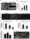
Figure 1. Taxol decreases scarring induced by spinal cord injury
(A) Representation of lesioned spinal cord (box). (B-C) Mid-saggital sections of lesion site from rats treated with (B) vehicle or (C) 256 ng/day 7 days post-injury. Scale bars, 300 μm. (D) Taxol significantly decreases fibrotic scarring (expressed as % of vehicle control; ** p= 0.002) without affecting wound size (E). Taxol does not affect glial compaction at 28 dpi (F). Data expressed as mean ± SEM.
Figure 2
Figure 2. Taxol dampens TGF-ß signalling
(A-B) In cultured astrocytes, Taxol counteracts the TGF-ß1 induced-nuclear translocation of Smad2/3 (arrowheads) causing cytoplasmic Smad2/3 accumulation (arrow). Results in (B) are mean ± SD (3 independent experiments, * p= 0.041). (C-F) Representative immunoblots. (C) Taxol treament increases total tubulin and decreases tyrosinated tubulin in the lesion site. (D) Kinesin-1 enrichment in microtubule fraction of Taxol-treated lesion site. (E) His-Smad2 binds to kinesin-1 of brain and spinal cord extracts. (F) Smad2 co-immunoprecipitates with kinesin-1. (G) Overlay of sequential binarized peroxisome images, color-coded by time as indicated, for a time series in which KIF5-FRB was recruited to peroxisomes upon addition of rapalog with (right) or without (left) 100 nM Taxol. Blue marks the initial distribution, whereas red marks regions targeted. Scale bars, 10 μm. (H) Time traces of the R90% (see online methods) KIF5-FRB and BICDN-FRB with or without 100 nM Taxol (D). Rapalog is added at 0:00 minutes. Time traces show average ± SEM of 5 (KIF5), 6 (KIF+Taxol), 3 (BICDN), and 8 (BICDN+Taxol) cells. (I) At 7 dpi, Taxol treatment interferes with the nuclear translocation of phospho-Smad 2/3 induced by SCI. Scale bar, 20 μm.
Figure 3
Figure 3. Taxol decreases meningeal cell migration and glycosaminoglycan release in vitro and in vivo
(A-B) 3 DIV, Taxol (1 and 10 nM) decreases meningeal cell migration induced by TGF-ß1. Scale bar, 300 μm; results in (B) are means ± SD from 3 independent experiments; * p= 0.003. (C) Taxol decreases fibronectin deposits induced by TGF-ß1 in meningeal cells. Scale bar, 300 μm. Taxol decreases GAGs released from control and TGF-ß1 stimulated meningeal cells (D) and astrocytes (E) . Results are means ± SD from 3 independent experiments; ** p= 0.008: * p=0.03). (F) Taxol treatment decreases the amount of GAGs induced by SCI. Results are means ± SEM, * p= 0.02. (G) Carbohydrate epitopes of CSPGs (CS-56) are abundant within the ECM in the control group. After Taxol treatment, CSPGs remain cytoplasmic. Scale bar, 75 μm.
Figure 4
Figure 4. Taxol promotes axonal regeneration and functional recovery
(A) Spinal cord horizontal sections of L4-L6 DRG axons labelled with CTB, 6 weeks after injury. Taxol treatment promotes regeneration of growth competent neurons (A, arrowheads). Scale bars, 50 μm. (B) Longest regenerating axon per animal ± SEM (** p= 0.002). (C) Spinal cord saggital sections stained with anti-5HT antibody 4 weeks after injury. After Taxol treatment, the caudal part of the cord is enriched in serotonergic fibers (C, arrowheads). Scale bars, 75 μm. (D-E) Quantification of 5HT-positive fibers caudal to the lesion ± SEM (*** p= 0.0001 (D) and ** p= 0.002 (E)). (F) Taxol treatment improves locomotor performance over the time.

Comment in

References

    1. Liu X, et al. PLoS Med. 2005 Dec;2:e354. - PMC - PubMed
    1. Westermann S, Weber K. Nat Rev Mol Cell Biol. 2003 Dec;4:938. - PubMed
    1. Erturk A, Hellal F, Enes J, Bradke F. J Neurosci. 2007 Aug 22;27:9169. - PMC - PubMed
    1. Witte H, Neukirchen D, Bradke F. J Cell Biol. 2008 Feb 11;180:619. - PMC - PubMed
    1. Klapka N, Muller HW. J Neurotrauma. 2006 Mar-Apr;23:422. - PubMed

Publication types

MeSH terms