. 2011 Feb 15;108(7):2759-64.
doi: 10.1073/pnas.1018823108. Epub 2011 Jan 27.

Primary cilia membrane assembly is initiated by Rab11 and transport protein particle II (TRAPPII) complex-dependent trafficking of Rabin8 to the centrosome

Affiliations

Primary cilia membrane assembly is initiated by Rab11 and transport protein particle II (TRAPPII) complex-dependent trafficking of Rabin8 to the centrosome

Christopher J Westlake et al. Proc Natl Acad Sci U S A. .

Abstract

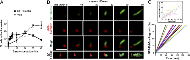
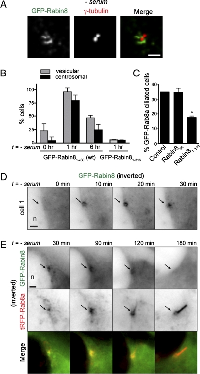
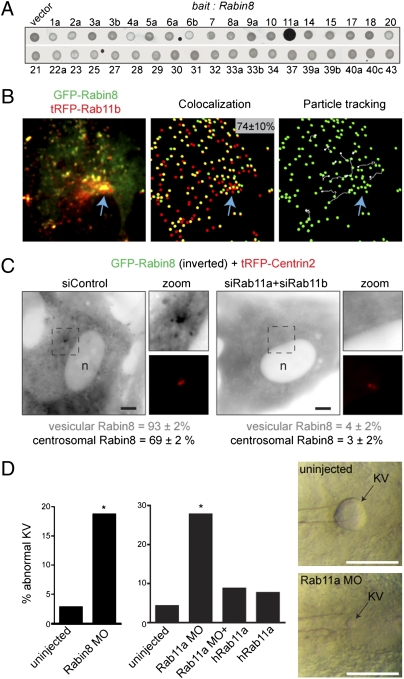
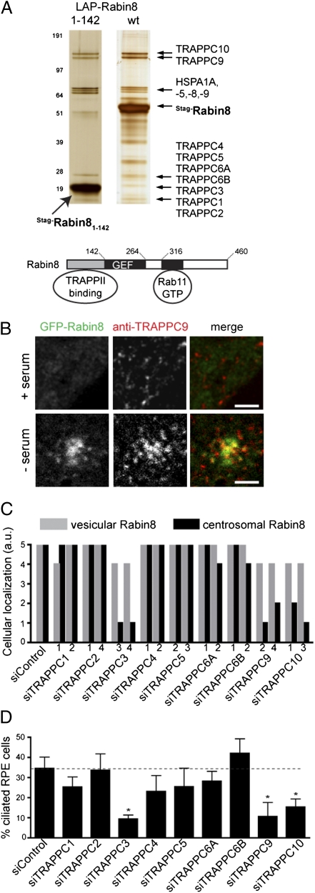
Sensory and signaling pathways are exquisitely organized in primary cilia. Bardet-Biedl syndrome (BBS) patients have compromised cilia and signaling. BBS proteins form the BBSome, which binds Rabin8, a guanine nucleotide exchange factor (GEF) activating the Rab8 GTPase, required for ciliary assembly. We now describe serum-regulated upstream vesicular transport events leading to centrosomal Rab8 activation and ciliary membrane formation. Using live microscopy imaging, we show that upon serum withdrawal Rab8 is observed to assemble the ciliary membrane in ∼100 min. Rab8-dependent ciliary assembly is initiated by the relocalization of Rabin8 to Rab11-positive vesicles that are transported to the centrosome. After ciliogenesis, Rab8 ciliary transport is strongly reduced, and this reduction appears to be associated with decreased Rabin8 centrosomal accumulation. Rab11-GTP associates with the Rabin8 COOH-terminal region and is required for Rabin8 preciliary membrane trafficking to the centrosome and for ciliogenesis. Using zebrafish as a model organism, we show that Rabin8 and Rab11 are associated with the BBS pathway. Finally, using tandem affinity purification and mass spectrometry, we determined that the transport protein particle (TRAPP) II complex associates with the Rabin8 NH(2)-terminal domain and show that TRAPP II subunits colocalize with centrosomal Rabin8 and are required for Rabin8 preciliary targeting and ciliogenesis.


Conflict of interest statement

Conflict of interest statement: C.J.W., K.J.W., K.E.E., L.P., C.C., D.S.K., R.H.S., and P.K.J. are employees of Genentech, Inc.

Figures

Fig. 1.
Fig. 1.
Rab8 localizes to developing cilia but departs mature cilia. (A) Rab8 is an early marker of ciliogenesis. GFP-Rab8a localization in cilia was scored in live RPE GFP-Rab8a cells, and Actub-positive cilia were counted in fixed cells (n = 75 cells). (B) Rab8 accumulates in the growing cilia. One hour after serum withdrawal, RPE GFP-Rab8a cells transiently expressing tRFP-Centrin2 for 24 h were imaged every 10 min by SDC microscopy over 6 h with concurrent imaging in both red and green channels. Representative contrast-adjusted images shown are projections of 20 z-sections (step size 0.5 μm). (Bottom Row) 3D surface rendering. (Scale bar: 5 μm.) (Movie S1). (C) Kinetics of ciliogenesis. Keying on the initiation of nascent cilia, GFP-Rab8a levels in 12 developing cilia were measured from projected z-stacks collected as described in B. Regression analysis was performed on raw data (Inset) to determine the time required for GFP fluorescence to reach maximal levels (106 ± 12 min).
Fig. 2.
Fig. 2.
Serum withdrawal triggers Rabin8 association with vesicles that accumulate dynamically at the centrosome before ciliary membrane assembly. (A) Rabin8 localizes to dynamic centrosomal vesicles following serum withdrawal. Serum-starved (60 min) RPE GFP-Rabin8 cells were fixed and stained with the centriolar marker γ-tubulin (Movie S2). (Scale bar: 2 μm.) (B) Serum starvation causes transient association of Rabin8 with membrane vesicles and accumulation at the centrosome mediated by its COOH-terminal domain. Shown is quantification of GFP-Rabin8 and GFP-Rabin81–316 (ΔC)-positive vesicles and centrosomal accumulation in RPE cells (n = 50) (Fig. S2 A and B). (C) The Rabin8 COOH-terminal domain is required for ciliation. GFP-Rab8a–positive cilia were counted in cells transiently expressing control (tRFP), tRFP-Rabin8, and tRFP-Rabin81–316 for 72 h; serum was removed for the last 24 h. GFP-positive cilia were counted only in tRFP-positive cells (n = 50; P < 0.001). (D) Time course of RPE cells accumulating GFP-Rabin8 at the centrosome after serum removal. Fluorescence signal (black) has been inverted in D and E for better viewing of structures. (Scale bar: 2 μm.) n, nucleus. (E) Rabin8 centrosomal targeting precedes Rab8a ciliary membrane formation and is reduced after ciliogenesis. RPE GFP-Rabin8 cells transiently expressing tRFP-Rab8a for 24 h were imaged live every 30 min after serum withdrawal. GFP and tRFP images were collected separately. (Scale bar: 2 μm.)
Fig. 3.
Fig. 3.
Rab11 is required for Rabin8 vesicle recruitment and centrosomal trafficking and is important for ciliated KV formation in zebrafish. (A) Rabin8 specifically interacts with the GTP-locked form of Rab11a (Q70L) by yeast two-hybrid analysis. Rabin8 was screened against a library of 37 constitutively active (“GTP-locked”) Rabs. One isoform for most of the 60 known Rabs was included (exceptions were Rab24, -26, -35, -36, and -38). (B) GFP-Rabin8 and tRFP-Rab11b vesicles colocalize and are transported to the centrosome. (Left) RPE GFP-Rabin8 cells transiently expressing tRFP-Rab11b (48 h) were serum starved for 1 h and imaged by live dual-view SDC microscopy. (Center) In five cells analyzed, 74 ± 10% of GFP-Rabin8 vesicles (green spheres) colocalized with tRFP-Rab11b structures (red spheres). (Right) Particle-tracking software was used to visualize the trajectory (shown in white) of Rab11b-Rabin8 double-positive vesicles. Blue arrows mark the pericentriolar region. (C) Rab11 is required for Rabin8 trafficking. Micrographs show representative siRNA-treated RPE GFP-Rabin8 cells expressing tRFP-Centrin2 that were serum-starved for 1 h. GFP-Rabin8 vesicle recruitment and centrosomal accumulation was scored in live cells (n = 50). Representative results from three independent experiments are shown. (Scale bar: 5 μm.) (D) Rabin8 and Rab11a knockdown caused defects in KV formation in zebrafish. Embryos were untreated or injected with 5 ng Rabin8 MO and 2.5 ng Rab11a MO as previously described (4). (Right) Micrographs show KVs from 8–10 somite embryos. (Left) Rabin8 MO caused KV abnormalities in 19% of embryos (29/155; P < 0.0001), compared with >4% of control embryos (3/108). (Center) Rab11 MO caused KV abnormalities in 27% (38/137; P < 0.0001) of embryos, compared with >4% of control embryos (3/71). Coinjection of hRab11a mRNA rescued the phenotype (9%, 11/126) in Rab11a MO-treated embryos (Fig. S3 J and K).
Fig. 4.
Fig. 4.
The TRAPPII complex associates with the Rabin8 NH2-terminal domain and is required for Rabin8 centrosomal targeting and primary cilium assembly. (A) LAP-tag purification of Rabin8-associated proteins. (Upper) LAP-Rabin8 and LAP-Rabin81–142 were purified by tandem affinity from 293Trex cells, and the eluted proteins were analyzed by SDS/PAGE (4–12% gradient) and silver stained; then 14 equally spaced gel slices were cut. TRAPPC components had high-percentage peptide coverage in gel slices analyzed by LC-MS/MS corresponding to their predicted molecular weight (Table S1). (Lower) Rabin8-binding domains important for centrosomal vesicular trafficking. (B) TRAPPC9 colocalizes with GFP-Rabin8 on centrosomal vesicles after serum starvation. Micrographs show RPE GFP-Rabin8 cells grown with or without serum for 60 min and fixed and stained with anti-TRAPPC9 antibodies before confocal laser scanning microscopy. (Whole-cell images are shown in Fig. S4E.) (Scale bars: 2 μm.) (C) TRAPPC3, -C9, and -C10 are required for Rabin8 centrosome trafficking. RPE GFP-Rabin8 cells expressing tRFP-Centrin2 were treated with two different ablating TRAPPC protein siRNAs for 72 h (knockdown efficiency is shown in Table S2) followed by serum starvation for 1 h. GFP-Rabin8 subcellular localization was scored in live cells (n = 50) by comparing siRab11a+siRab11b-treated cells (score = 0; Fig. 3C) with siControl-treated cells (score = 5). Results from three independent experiments are shown (Fig. S4H). (D) TRAPPC3, -C9, and -C10 are required for ciliation. RPE cells were treated with siRNAs as in C, serum starved 24 h, and stained with Actub and pericentrin antibodies (n = 75 cells; P < 0.0001). Representative data from three independent experiments are shown.

References

    1. Scholey JM, Anderson KV. Intraflagellar transport and cilium-based signaling. Cell. 2006;125:439–442. - PubMed
    1. Hattula K, Furuhjelm J, Arffman A, Peränen J. A Rab8-specific GDP/GTP exchange factor is involved in actin remodeling and polarized membrane transport. Mol Biol Cell. 2002;13:3268–3280. - PMC - PubMed
    1. Nachury MV, et al. A core complex of BBS proteins cooperates with the GTPase Rab8 to promote ciliary membrane biogenesis. Cell. 2007;129:1201–1213. - PubMed
    1. Yen HJ, et al. Bardet-Biedl syndrome genes are important in retrograde intracellular trafficking and Kupffer's vesicle cilia function. Hum Mol Genet. 2006;15:667–677. - PubMed
    1. Sorokin S. Centrioles and the formation of rudimentary cilia by fibroblasts and smooth muscle cells. J Cell Biol. 1962;15:363–377. - PMC - PubMed

Publication types