. 2011 Apr;300(4):G627-36.
doi: 10.1152/ajpgi.00563.2010. Epub 2011 Jan 27.

Epidermal growth factor upregulates serotonin transporter in human intestinal epithelial cells via transcriptional mechanisms

Affiliations

Epidermal growth factor upregulates serotonin transporter in human intestinal epithelial cells via transcriptional mechanisms

Ravinder K Gill et al. Am J Physiol Gastrointest Liver Physiol. 2011 Apr.

Abstract

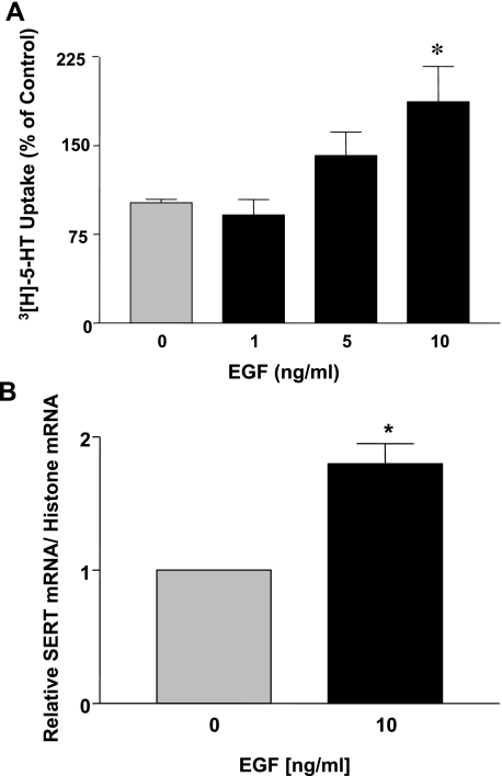
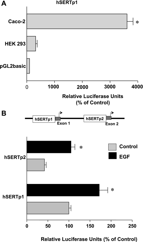
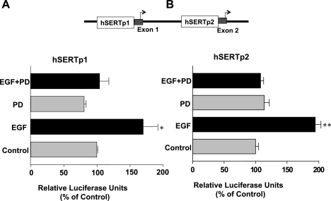
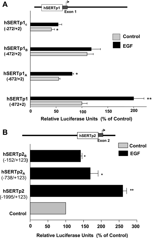
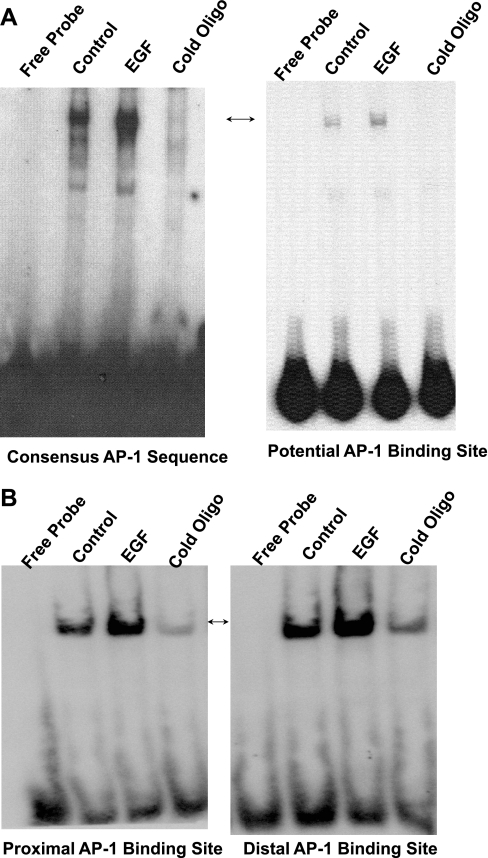
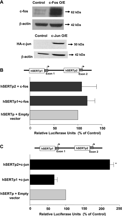
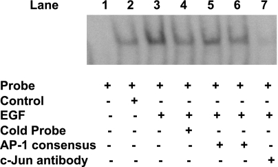
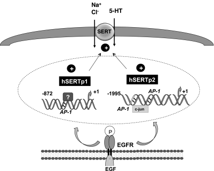
Serotonin transporter (SERT) regulates extracellular availability of serotonin and is a potential pharmacological target for gastrointestinal disorders. A decrease in SERT has been implicated in intestinal inflammatory and diarrheal disorders. However, little is known regarding regulation of SERT in the intestine. Epidermal growth factor (EGF) is known to influence intestinal electrolyte and nutrient transport processes and has protective effects on intestinal mucosa. Whether EGF regulates SERT in the human intestine is not known. The present studies examined the regulation of SERT by EGF, utilizing Caco-2 cells grown on Transwell inserts as an in vitro model. Treatment with EGF from the basolateral side (10 ng/ml, 24 h) significantly stimulated SERT activity (∼2-fold, P < 0.01) and mRNA levels compared with control. EGF increased the activities of the two alternate promoter constructs for human SERT gene: SERT promoter 1 (hSERTp1, upstream of exon 1a) and SERT promoter 2 (hSERTp2, upstream of exon 2). Inhibition of EGF receptor (EGFR) tyrosine kinase activity by PD168393 (1 nM) blocked the stimulatory effects of EGF on SERT promoters. Progressive deletions of the SERT promoter indicated that the putative EGF-responsive elements are present in the -672/-472 region of the hSERTp1 and regions spanning -1195/-738 and -152/+123 of hSERTp2. EGF markedly increased the binding of Caco-2 nuclear proteins to the potential AP-1 cis-elements present in EGF-responsive regions of hSERTp1 and p2. Overexpression of c-jun but not c-fos specifically transactivated hSERTp2, with no effects on hSERTp1. Our findings define novel mechanisms of transcriptional regulation of SERT by EGF via EGFR at the promoter level that may contribute to the beneficial effects of EGF in gut disorders.


Figures

Fig. 1.
Fig. 1.
Epidermal growth factor (EGF) treatment of Caco-2 cells increases serotonin transporter (SERT) function and expression. Caco-2 cells grown on Transwell inserts were treated with EGF (1–10 ng/ml, 24 h) from the basolateral side in serum-free Caco-2 medium supplemented with 0.1% bovine serum albumin (BSA). A: SERT function was measured as [3H]serotonin ([3H]5-HT) uptake from the apical side. Data are shown as % of control (means ± SE of values obtained from at least 3 different experiments performed in triplicate). Absolute mean control value: 0.57 pmol·mg protein−1·5 min−1. *P < 0.01 compared with control. B: total RNA was extracted, and quantitative real-time RT-PCR was performed with SYBR Green fluorescent dye. Human SERT (hSERT) mRNA levels were normalized to the levels of histone mRNA. Data are shown as means ± SE of values obtained from at least 4 different experiments performed in triplicate. *P < 0.01 compared with control.
Fig. 2.
Fig. 2.
Effects of EGF on hSERT promoter activities in Caco-2 cells. A: hSERTp1 is active in Caco-2 cells. Caco-2 and HEK-293 cells were transiently cotransfected with the hSERTp1 promoter construct along with the β-galactosidase mammalian expression vector. Promoter activity was measured by firefly luciferase assay and represented as relative luciferase units normalized to β-galactosidase activity to adjust for transfection efficiency. The activity of the pGL2 empty vector alone in Caco-2 cells or HEK-293 cells is represented as 100%. hSERTp1 was highly active in Caco-2 cells. Results were obtained from 3 separate experiments and are expressed as means ± SE. *P < 0.0001 compared with empty vector. B: effect of EGF on activity of hSERTp1 and hSERTp2. Caco-2 cells were transiently cotransfected with hSERTp1 or hSERTp2 fragments along with β-galactosidase vector to adjust for transfection efficiency. Twenty-four hours after transfection, cells were incubated with 10 ng/ml EGF for another 24 h. hSERTp1 exhibited higher activity than hSERTp2 in Caco-2 cells. EGF increased the activity of both hSERTp1 and hSERTp2. Data are represented as % of control and represent means ± SE of 5 or 6 different experiments performed in triplicate. *P < 0.05 compared with control.
Fig. 3.
Fig. 3.
Stimulation of hSERTp1 and hSERTp2 is EGF receptor (EGFR) dependent. Caco-2 cells were transfected with hSERTp1 (A) or hSERTp2 (B) fragments along with β-galactosidase vector. Twenty-four hours after transfection, cells were pretreated from the basolateral side with the cell-permeant EGFR tyrosine kinase activity inhibitor PD168393 (PD, 1 nM) for 1 h, followed by coincubation with EGF (10 ng/ml) for another 24 h. SERT promoter activities were assessed by luciferase assays normalized to β-galactosidase activity. Values represent % of control derived from means ± SE of 3 different experiments. *P < 0.05, **P < 0.01 compared with respective controls.
Fig. 4.
Fig. 4.
EGF-responsive regions of hSERTp1 and hSERTp2. A: hSERTp1: progressive 5′ deletions of hSERTp1 treated with EGF. Different promoter constructs of hSERTp1 were treated with EGF (10 ng/ml, 24 h). Twenty-four hours after treatment, cells were harvested for measurement of promoter activity by luciferase assay. Values were normalized to β-galactosidase to adjust for transfection efficiency. The stimulatory effect of EGF was abolished on deletion to −472/+2 bp. Data were obtained from at least 5 different experiments performed in triplicate and are shown as means ± SE. Results are expressed as percentages of control. *P < 0.05, **P < 0.001 compared with control. B: hSERTp2: progressive 5′ deletion constructs of hSERTp2 were cotransfected in Caco-2 cells with β-galactosidase vector, and effect of EGF on promoter activity was measured. EGF-responsive region predominantly spans −1995/−738 region of hSERTp2. Data were obtained from at least 3 different experiments performed in triplicate and are shown as means ± SE. Results are expressed as % of control. *P < 0.01, **P < 0.001 compared with control.
Fig. 5.
Fig. 5.
Effect of EGF on nuclear protein binding to potential AP-1 cis-elements of hSERTp1 (A) and hSERTp2 (B). A: 8 μg of nuclear extracts from control (untreated) or EGF-treated (10 ng/ml, 24 h) Caco-2 cells was incubated with the 32P-labeled AP-1 cis-element of hSERTp1 (right) or with 32P-labeled consensus AP-1 sequence (left). Protein-DNA complexes competed in the presence of excess of cold unlabeled oligonucleotide probe. B: nuclear extracts from control (untreated) or EGF-treated (10 ng/ml, 24 h) Caco-2 cells were incubated with digoxigenin (DIG)-labeled AP-1 potential cis-element of hSERTp2. Binding of both the potential AP-1 sites located proximal to the transcription site (left) and the distal AP-1 site (right) is shown. Protein-DNA complexes competed in the presence of excess of cold unlabeled oligonucleotide probe, showing specificity of binding. Representative gels of 3 or 4 separate experiments with similar results are shown.
Fig. 6.
Fig. 6.
Effect of c-fos or c-jun overexpression on hSERTp1 and hSERTp2 activities. A: expression of c-jun or c-fos. Cells were cotransfected with hSERT promoter constructs and mammalian expression vectors for c-fos or hemagglutinin (HA)-tagged c-jun along with the β-galactosidase vector. Cells were harvested after 24 h, and protein lysates were run on SDS-PAGE, followed by immunoblotting with anti-c-fos or anti-HA antibodies to examine the protein expression of c-fos and c-jun, respectively. β-Actin was used as an internal control. O/E, overexpressing. Data were obtained from 3 separate experiments. B: effect of c-fos overexpression on hSERTp1 and hSERTp2 activities. Caco-2 cells were transiently transfected with c-fos mammalian expression vector for 24 h along with promoter construct hSERTp1 or hSERTp2. Control cells were transfected with empty vector for c-fos (PCDN3.1) and hSERTp1 or hSERTp2. Promoter activity was measured by luciferase assay normalized to β-galactosidase to adjust for transfection efficiency. c-fos overexpression failed to alter the activity of hSERTp1 or hSERTp2 compared with respective controls. Data are % of respective controls represented as means ± SE of 5 individual experiments performed in triplicate. C: c-jun overexpression transactivates hSERTp2 activity. Caco-2 cells were transiently transfected with c-jun mammalian expression vector for 24 h along with promoter construct hSERTp1 or hSERTp2. Control cells were cotransfected with empty vector and hSERTp1 or hSERTp2. c-jun overexpression transactivated hSERTp2, with no effect on hSERTp1 compared with their respective controls. Data are represented as % of control represented as means ± SE of 3 individual experiments performed in triplicate. *P < 0.001 compared with control.
Fig. 7.
Fig. 7.
c-jun binds to potential AP-1 cis-elements of hSERTp2. Nuclear extracts from control (untreated) or EGF-treated (10 ng/ml, 24 h) Caco-2 cells were incubated with DIG-labeled distal AP-1 cis-element of the hSERTp2. DNA-protein complexes in response to EGF treatment were competed by excess of unlabeled probe (lane 4). Excess of cold oligonucleotides representing the AP-1 consensus sequence (125-fold excess, lane 5; 150-fold excess, lane 6) competed out the increase in binding observed in EGF-treated nuclear extracts (lane 3). Addition of c-jun antibody abolished the increase in binding in response to EGF treatment (lane 7). These data indicate that c-jun binds to the potential AP-1 cis-elements of hSERTp2. A representative blot is shown.
Fig. 8.
Fig. 8.
Schematic of the proposed model of EGF-mediated effects on SERT.

Similar articles

Cited by

References

    1. Alrefai WA, Annaba F, Sarwar Z, Dwivedi A, Saksena S, Singla A, Dudeja PK, Gill RK. Modulation of human Niemann-Pick C1-like 1 gene expression by sterol: role of sterol regulatory element binding protein 2. Am J Physiol Gastrointest Liver Physiol 292: G369–G376, 2007 - PubMed
    1. Banan A, Zhang LJ, Shaikh M, Fields JZ, Farhadi A, Keshavarzian A. Key role of PLC-gamma in EGF protection of epithelial barrier against iNOS upregulation and F-actin nitration and disassembly. Am J Physiol Cell Physiol 285: C977–C993, 2003 - PubMed
    1. Berger M, Gray JA, Roth BL. The expanded biology of serotonin. Annu Rev Med 60: 355–366, 2009 - PMC - PubMed
    1. Bishop WP, Wen JT. Regulation of Caco-2 cell proliferation by basolateral membrane epidermal growth factor receptors. Am J Physiol Gastrointest Liver Physiol 267: G892–G900, 1994 - PubMed
    1. Borthakur A, Gill RK, Tyagi S, Koutsouris A, Alrefai WA, Hecht GA, Ramaswamy K, Dudeja PK. The probiotic Lactobacillus acidophilus stimulates chloride/hydroxyl exchange activity in human intestinal epithelial cells. J Nutr 138: 1355–1359, 2008 - PMC - PubMed

Publication types

MeSH terms

Substances