. 2011 May;300(5):G749-60.
doi: 10.1152/ajpgi.00306.2010. Epub 2011 Jan 27.

Muscarinic receptor agonists stimulate human colon cancer cell migration and invasion

Affiliations

Muscarinic receptor agonists stimulate human colon cancer cell migration and invasion

Angelica Belo et al. Am J Physiol Gastrointest Liver Physiol. 2011 May.

Abstract

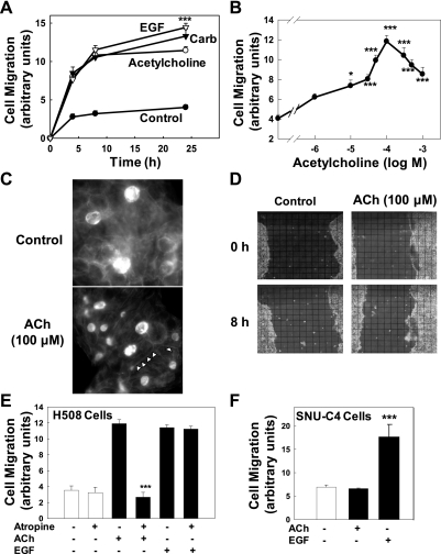
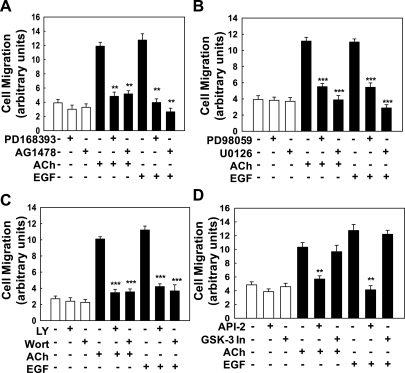
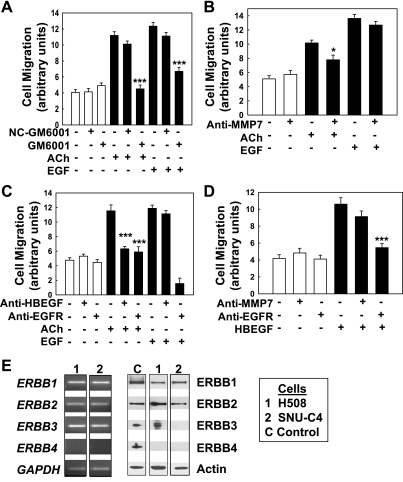
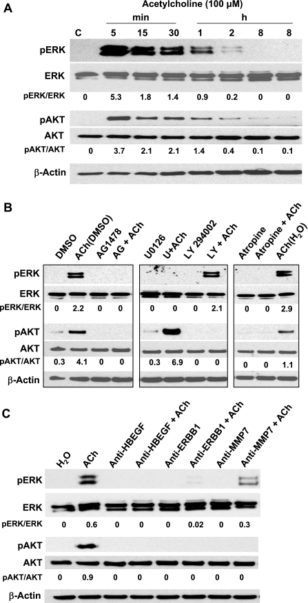
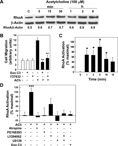
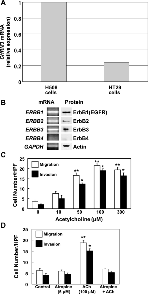
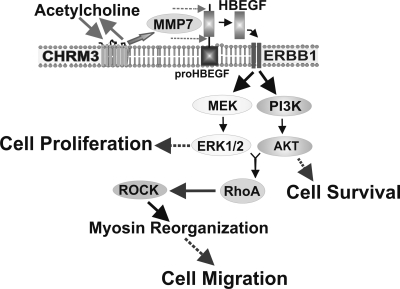
Muscarinic receptors (CHRM) are overexpressed in colon cancer. To explore a role for muscarinic receptor signaling in colon cancer metastasis, we used human H508 and HT29 colon cancer cells that coexpress epidermal growth factor (ERBB) and CHRM3 receptors. In a wound closure model, following 8-h incubation of H508 cells with 100 μM ACh we observed a threefold increase in cell migration indistinguishable from the actions of epidermal growth factor (EGF). Atropine blocked the actions of ACh but not of EGF. In SNU-C4 colon cancer cells that express ERBB but not CHRM, EGF caused a threefold increase in migration; ACh had no effect. ACh-induced cell migration was attenuated by chemical inhibitors of ERBB1 activation, by anti-ERBB1 antibody, and by inhibitors of ERK and phosphatidylinositol 3-kinase (PI3K) signaling. Consistent with matrix metalloproteinase-7 (MMP7)-mediated release of an ERBB1 ligand, heparin binding epidermal growth factor-like growth factor (HBEGF), ACh-induced migration was inhibited by an MMP inhibitor and by anti-MMP7 and -HBEGF antibodies. ACh-induced cell migration was blocked by inhibiting RhoA and ROCK, key proteins that interact with the actin cytoskeleton. ACh-induced RhoA activation was attenuated by agents that inhibit ERBB1, ERK, and PI3K activation. Collectively, these findings indicate that ACh-induced cell migration is mediated by MMP7-mediated release of HBEGF, an ERBB ligand that activates ERBB1 and downstream ERK and PI3K signaling. In a cell invasion model, ACh-induced HT29 cell invasion was blocked by atropine. In concert with previous observations, these findings indicate that muscarinic receptor signaling plays a key role in colon cancer cell proliferation, survival, migration, and invasion.


Figures

Fig. 1.
Fig. 1.
Acetylcholine (ACh) stimulates colon cancer cell migration in a time- and concentration-dependent manner. Human colon cancer cells were plated at confluence before a linear “wound” was made. As described in materials and methods, photomicrographs were taken immediately after wounding, before test agents were added, and again as described below. A: time course for effects of ACh (100 μM), epidermal growth factor (EGF; 10 μg/ml), and carbamylcholine (Carb; 100 μM) on H508 colon cancer cell migration. Cell migration was measured at the time points indicated. B: dose-response curve for the effects of ACh on H508 cell migration. Plates were incubated with the indicated concentrations of ACh and cell migration was measured at 8 h. The data point on the vertical axis represents cell migration without ACh. C: representative photographs showing that incubation with ACh (100 μM for 8 h) induces stress fiber formation in H508 cells. Arrowheads in bottom image indicate representative stress fibers in ACh-treated cells. D: representative photographs showing cell culture wounding assay immediately after scraping with pipette tip (0 h) and 8 h after incubation with ACh (100 μM) or vehicle (control). The overlying grid used to measure cell migration is shown. E: preincubation with atropine (1 μM) blocked ACh (100 μM)-induced but not EGF (10 μg/ml)-induced H508 cell migration. Cell migration was measured 8 h after addition of test agents. F: in SNU-C4 human colon cancers that do not express muscarinic receptors, cell migration, measured 8 h after addition of test agents, was stimulated by EGF (10 μg/ml) but not ACh (100 μM). Values in A, B, D, and E are means ± SE from at least 3 separate experiments. *, ***P < 0.05 and 0.001, respectively, compared with control (treatment with vehicle alone).
Fig. 2.
Fig. 2.
ACh-induced colon cancer cell migration is dependent on ERBB1 activation and post-ERBB1 signaling. Human colon cancer cells were plated at confluence before a linear wound was made. As described in materials and methods, photomicrographs were taken immediately after a linear wound was made and again 8 h after addition of test agents. A: ACh (100 μM)- and EGF (10 μg/ml)-induced migration of H508 colon cancer cells was attenuated by adding ERBB1 activation inhibitors (PD168393, AG1478; both 10 μM). B: ACh (100 μM)- and EGF (10 μg/ml)-induced migration of H508 colon cancer cells was attenuated by adding ERK activation inhibitors (PD98059 and U0126; both 10 μM). C: ACh (100 μM)- and EGF (10 μg/ml)-induced migration of H508 colon cancer cells was attenuated by adding phosphatidylinositol 3-kinase (PI3K) activation inhibitors [10 μM LY294002 (LY) and 50 nM wortmannin (Wort)]. D: ACh (100 μM)- and EGF (10 μg/ml)-induced migration of H508 colon cancer cells was attenuated by adding an inhibitor of AKT activation (5 μM API-2) but not by adding a GSK-3 inhibitor (50 nM GSK-3 inhibitor IX). Values are means ± SE from at least 3 separate experiments. **, ***P < 0.01 and 0.001, respectively, compared with control (treatment with vehicle alone).
Fig. 3.
Fig. 3.
ACh-induced colon cancer cell migration is mediated by activation of matrix metalloproteinases and release of an ERBB ligand. Human colon cancer cells were plated at confluence before a linear wound was made. As described in materials and methods, photomicrographs were taken immediately after a linear wound was made and again 8 h after adding test agents. A: a broad-spectrum matrix metalloproteinase inhibitor (10 μM GM6001) abolished ACh-induced H508 cell migration. GM6001 also attenuated EGF-induced cell migration. In contrast, a structurally similar control agent (10 μM NC-GM6001) did not alter ACh- or EGF-induced cell migration. B: anti-MMP7 antibody (5 μg/ml) attenuated the actions of ACh but did not alter EGF-induced cell migration. C: anti-HBEGF (1 μg/ml) and anti-ERBB1 antibodies attenuated ACh-induced cell migration. Whereas anti-ERBB1 antibody blocked the actions of EGF, anti-HBEGF antibody did not alter EGF-induced cell migration. D: recombinant HBEGF (20 ng/ml) stimulated H508 cell migration. These actions were blocked by anti-EGFR antibody (0.02 μg/ml) but not by anti-MMP7 antibody (5 μg/ml). E: expression of ERBB subtypes in human colon cancer cells. Left: quantitative real-time PCR (q-PCR) demonstrates expression of mRNA for ERBB subtypes in H508 and SNU-C4 human colon cancer cells. Right: immunoblots demonstrate expression of protein for ERBB subtypes in lysates of H508 and SNU-C4 cells. Values in AD are means ± SE from at least 3 separate experiments. *, ***P < 0.05 and 0.001, respectively, compared with control (treatment with vehicle alone).
Fig. 4.
Fig. 4.
In H508 colon cancer cells, ACh activates ERBB1-dependent ERK and PI3K signaling. ERK and AKT activation (phosphorylation) was determined by immunoblotting proteins from H508 cell lysates as described in materials and methods. A: representative immunoblot showing that ACh (100 μM) stimulates time-dependent ERK and AKT phosphorylation (pERK and pAKT) but no change in expression of total ERK and AKT. Densitometry was used to compare signals for phosphorylated (activated) compared with total proteins. B: representative immunoblot exploring upstream signaling pathways that mediate ACh-induced AKT and ERK activation. ACh-induced phosphorylation of AKT is attenuated by inhibitors of ERBB1 (10 μM AG1478) and PI3K (10 μM LY294002), but not by an ERK inhibitor (10 μM U0126). ACh-induced phosphorylation of ERK is attenuated by ERBB1 and ERK inhibitors, but not by a PI3K inhibitor. ACh-induced activation of both AKT and ERK was blocked by atropine (5 μM), a muscarinic receptor inhibitor. DMSO (0.5%), a solvent for U0126 and LY294002, increased ACh-induced AKT phosphorylation but did not alter ACh-induced ERK phosphorylation. C: representative immunoblot showing that in H508 cells, ACh-induced ERK and AKT activation (phosphorylation) was abolished or attenuated by anti-HBEGF, anti-ERBB1 and anti-MMP7 antibodies. For each condition, the ratio of signal intensity for activated ERK and AKT, compared with that for total ERK and AKT, respectively, was calculated. β-Actin was used as a loading control. All immunoblots are representative of at least 3 separate experiments. C, control (treatment with vehicle alone).
Fig. 5.
Fig. 5.
Actions of ACh on expression and activation of RhoA and ROCK. A: in H508 colon cancer cells, incubation with 100 μM ACh for up to 8 h did not alter expression of RhoA. RhoA expression was determined by immunoblotting proteins from H508 cell lysates after incubation for the times indicated. For each time point, the ratio of signal intensity for RhoA compared with that for β-actin (loading control) was calculated. Immunoblots shown are representative of 3 others. B: chemical inhibitors of RhoA [exoenzyme C3 (ExoC3), 5 μg/ml] and ROCK (Y27632, 75 μM) abolished ACh-induced H508 cell migration. After preincubation with inhibitors, cells were plated at confluence before a linear wound was made. Cells were incubated with or without ACh (100 μM) for an additional 8 h. As described in materials and methods, photomicrographs were taken immediately after a linear wound was made and again 8 h after addition of ACh. **P < 0.01 compared with ACh alone. Cell migration with ACh plus ExoC3 and Y27632 was not significantly different than control migration with vehicle alone. C: time course for ACh-induced activation of RhoA. In H508 cells, RhoA activation was measured using an ELISA-based assay (materials and methods) before (time = 0 min) and at the indicated times after adding ACh (solid bars). Results are expressed as a percentage of the maximal response obtained with 100 μM ACh (5 min). *P < 0.05 compared with basal RhoA activation (0 min). D: ACh-induced RhoA activation was blocked by inhibitors of muscarinic receptor, ERBB1, ERK, and PI3K signaling. After 5 min incubation with test agents, RhoA activation was measured as described in materials and methods. Bar graph shows effects of adding water-soluble (atropine, ExoC3) and methanol-soluble (PD168393, LY294002, U0126) inhibitors to 100 μM ACh. Vehicle (water and 0.5% methanol), atropine (5 μM), and ExoC3 (5 μg/ml) were used as controls. Basal RhoA activation was not altered by PD168393, LY294002, and U0126 (all 10 μM; data not shown) but these inhibitors all attenuated ACh-induced RhoA activation. ***P < 0.001 for 100 μM ACh compared with vehicle alone (either water or 0.5% methanol). Values shown in bar graphs (BD) represent means ± SE from at least 3 separate experiments.
Fig. 6.
Fig. 6.
Actions of ACh on HT29 human colon cancer cell migration and invasion. A: relative expression of CHRM3 mRNA in HT29 and H508 colon cancer cells. B: expression of ERBB subtypes in HT29 cells. Left: q-PCR demonstrates expression of mRNA for ERBB subtypes. Right: immunoblots demonstrate expression of protein for ERBB subtypes in HT29 cell lysates. C: ACh stimulates dose-dependent increases in HT29 cell migration and invasion. Cell migration and invasion assays were performed by using BD Biocoat Invasion Chambers without or with the Matrigel insert, respectively. HPF, high-powered field. D: HT29 cell migration and invasion stimulated by 100 μM ACh is blocked by preincubation with 5 μM atropine. Values shown in bar graphs (C and D) represent means ± SE from at least 3 separate experiments. *, **P < 0.05 and 0.01, respectively, compared with control (treatment with vehicle alone). q-PCR primer pairs for experiments in A and B are shown in Table 1.
Fig. 7.
Fig. 7.
Cartoon illustrating proposed intracellular mechanisms whereby muscarinic receptor activation stimulates human colon cancer cell migration. Muscarinic receptor ligands (e.g., ACh) bind to type 3 muscarinic receptors (CHRM3) on H508 cells, thereby activating matrix metalloproteinase-7 (MMP7) with consequent release of an ERBB receptor ligand (HBEGF). Published work indicates that, downstream of ERBB1 activation, ERK activation stimulates colon cancer cell proliferation and PI3K/AKT activation augments cell survival (5, 7, 8, 26, 35, 41). Novel findings reported in this communication include the observation that post-ERBB1 signaling by both the ERK and PI3K cascades stimulates RhoA activation and colon cancer cell migration.

Similar articles

Cited by

References

    1. Amado RG, Wolf M, Peeters M, Van Cutsem E, Siena S, Freeman DJ, Juan T, Sikorski R, Suggs S, Radinsky R, Patterson SD, Chang DD. Wild-type KRAS is required for panitumumab efficacy in patients with metastatic colorectal cancer. J Clin Oncol 26: 1626–1634, 2008 - PubMed
    1. Antonyak MA, Li B, Regan AD, Feng Q, Dusaban SS, Cerione RA. Tissue transglutaminase is an essential participant in the epidermal growth factor-stimulated signaling pathway leading to cancer cell migration and invasion. J Biol Chem 284: 17914–17925, 2009 - PMC - PubMed
    1. Balak MN, Gong Y, Riely GJ, Somwar R, Li AR, Zakowski MF, Chiang A, Yang G, Ouerfelli O, Kris MG, Ladanyi M, Miller VA, Pao W. Novel D761Y and common secondary T790M mutations in epidermal growth factor receptor-mutant lung adenocarcinomas with acquired resistance to kinase inhibitors. Clin Cancer Res 12: 6494–6501, 2006 - PubMed
    1. Belo A, Cheng K, Shant J, Raufman JP. Acetylcholine-induced colon cancer cell migration is mediated by epidermal growth factor receptor (EGFR)-dependent activation of both ERK and PI3K signaling (Abstract). Gastroenterology 136: S2040, 2009
    1. Cheng K, Raufman JP. Bile acid-induced proliferation of a human colon cancer cell line is mediated by transactivation of epidermal growth factor receptors. Biochem Pharmacol 70: 1035–1047, 2005 - PubMed

Publication types

MeSH terms