. 2011 May 2;52(6):2930-7.
doi: 10.1167/iovs.10-5940.

Smad4 is required predominantly in the developmental processes dependent on the BMP branch of the TGF-β signaling system in the embryonic mouse retina

Affiliations

Smad4 is required predominantly in the developmental processes dependent on the BMP branch of the TGF-β signaling system in the embryonic mouse retina

Deepa Murali et al. Invest Ophthalmol Vis Sci. .

Abstract

Purpose: The present study was aimed at defining developmental roles of Smad4, a key mediator of the TGF-β superfamily signaling system, in the embryonic mouse retina.

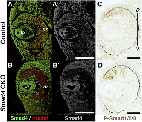
Methods: Using a Cre/loxP-mediated conditional gene targeting approach, Smad4 gene function was deleted from the embryonic mouse retina. Mutant phenotypes were morphologically and molecularly examined.

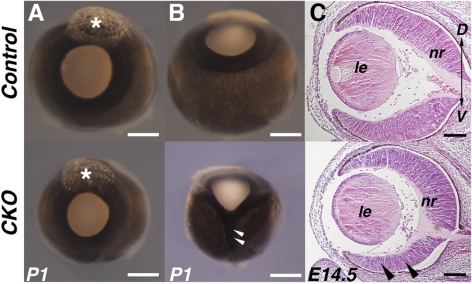
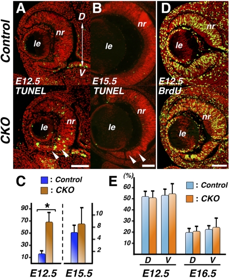
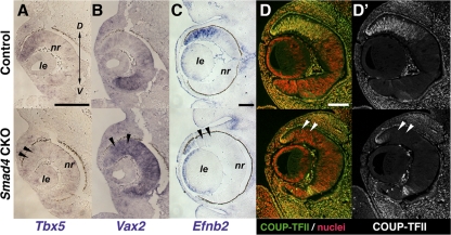
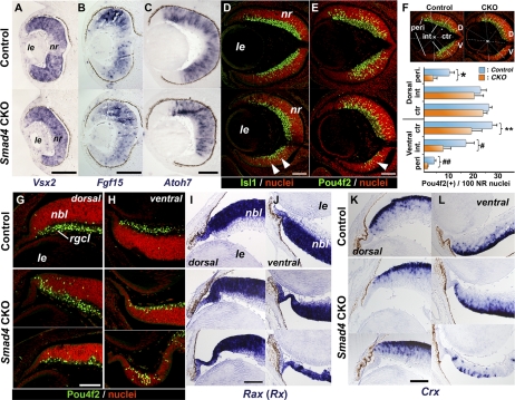
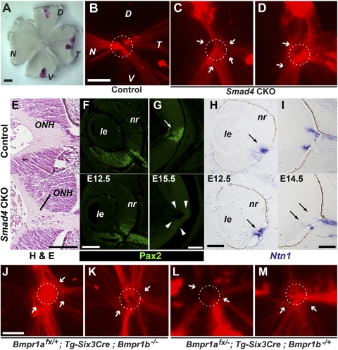
Results: Loss of Smad4 in the developing retina led to varying degrees of microphthalmia at birth, presumably because of elevated apoptosis observed transiently at embryonic day 12.5 in the developing retina. This was also associated with an apparent delay in accumulation of retinal ganglion cells. Smad4 conditional mutants also exhibited alterations of retinal spatial patterning along the dorsal-ventral axis, consistent with a known function of BMP signaling in the embryonic retina. However, despite a known role for BMP signaling in retinal cell survival, proliferation, and differentiation, Smad4 mutant retinal progenitor cells were capable of maintaining growth and neurogenesis throughout embryonic development. We also found that the loss of Smad4 led to abnormal targeting of retinal ganglion cell axons to the optic nerve head, a phenotype consistent with reduced BMP signaling in the developing retina.

Conclusions: These results suggest that Smad4 is essential for a subset of, but not all, TGF-β/BMP-dependent developmental processes in the embryonic retina. In addition, genetic requirements for Smad4 in the embryonic retina are evident predominantly in the developmental events regulated by the BMP branch of the TGF-β signaling pathway.


Figures

Figure 1.
Figure 1.
Microphthalmia and ventral retinal thinning in Smad4-CKO mutants. (A, B) Newborn eyes from control (top) and Smad4-CKO mutants (bottom) viewed from anterior (A) and ventral (B) orientations. The mutant eye (A) is a representative of mild microphthalmia, whereas a more severe case with concomitant coloboma is shown (B). Asterisks: resin marking dorsal side of the eye. (C) Embryonic eyes sectioned through the dorsal-ventral axis showing apparent thinning in the ventral quadrant of the mutant retina (arrowheads). D, dorsal; le, lens; nr, neural retina; V, ventral. Scale bars: 1 mm (A, B); 100 μm (C).
Figure 2.
Figure 2.
Embryonic retinal growth in Smad4-CKO mutants. (A) TUNEL staining on coronal sections showing elevated apoptotic cell death predominantly in the ventral retina of a Smad4-CKO mutant at E12.5 (arrowheads). (B) Later in development at E15.5, the Smad4-CKO retina no longer shows evidence of increased apoptosis, even in the thinned ventral retina (arrowheads). (C) Quantification of TUNEL-positive nuclei (per 1000 retinal cells) at E12.5 (n = 6 for control; n = 8 for mutants; *P = 0.0045) and E15.5 (n = 4 for control; n = 6 for mutants). (D) Representative images from BrdU incorporation assays at E12.5. (E) Quantification of BrdU incorporation in the dorsal (D) and ventral (V) regions of the developing retinas at E12.5 (n = 4) and E16.5 (n = 6). No statistically significant differences between controls and mutants are detected. D, dorsal; le, lens; nr, neural retina; V, ventral. Scale bars, 100 μm.
Figure 3.
Figure 3.
Expression of Smad4 and phospho-Smad1/5/8 in the developing retina. (A, B′) Immunohistochemical detection of Smad4 in the developing retina at E11.5, revealing wide distribution of Smad4 in and around the eye, including the retina, in the control (A, A′). In a CKO littermate, Smad4 immunoreactivity is diminished specifically in the retina (B, B′). (C, D) Immunolocalization of phospho-Smad1/5/8 in the embryonic retina at E12.5, revealing high levels of signal in the dorsal compartment in the CKO mutant (D), comparable to the control (C). D, dorsal; le, lens; nr, neural retina; V, ventral. Scale bars, 100 μm.
Figure 4.
Figure 4.
Defects in dorsal-ventral retinal patterning in Smad4-CKO mutants. (A, B) Expression of the dorsal marker Tbx5 (A) and ventral marker Vax2 (B) in E11.5 retinas. Arrowheads: loss of dorsal Tbx5 expression (A, bottom) and dorsal expansion of Vax2 expression domain (B, bottom), respectively. (C) Expression of Efnb2 in E14.5 retinas, showing diminished expression in the CKO mutant (arrowheads). (D, D′) Expression of COUP-TFII in E13.5 retinas. Although normally localized dorsally in the developing normal retina (top), its expression is lost in the Smad4-CKO retina (bottom, arrowheads). D, dorsal; le, lens; nr, neural retina; V, ventral. Scale bars, 100 μm.
Figure 5.
Figure 5.
Expression of markers for retinal growth and differentiation in Smad4-CKO mutants. (A, B) Expression of Vsx2 (Chx10) at E11.5 (A) and Fgf15 at E12.5 (B), both implicated in retinal progenitor growth. (C) Expression of the proneural gene Atoh7 (Math5) in the developing retina at E12.5. (D, E) Distribution of differentiating RGC markers Isl1 (D) and Pou4f2 (Brn3b) (E) in the developing retina at E14.5. (F) Regional distribution of Pou4f2-expressing cells in E14.5 retinas. Dorsal and ventral retinas in reference to the central depression of the ONH were, respectively, divided into peripheral (peri), intermediate (int), and central (ctr) domains. These domains were demarcated in equal angles from the reference point (X) between the distal tip of the retina and the ONH. The reference point was defined as the midpoint between the distal tip of the lens and the lumen of the ONH. Numbers of RGCs were counted separately in each of these domains of the neural retina (n = 4 for control; n = 6 for mutants; *P = 0.011; **P = 0.046; #P = 0.029; ##P = 0.0037). (GL) Expression of Pou4f2 (G, H), Rax (I, J), and Crx (K, L) in peripheral regions of the dorsal (G, I, K) and ventral (H, J, L) retinas at E18.5. For Smad4-CKO mutants, moderately thinned and more severely affected examples are shown in the middle and bottom rows, respectively. D, dorsal; le, lens; nbl, neuroblastic layer; nr, neural retina; rgcl, RGC layer; V, ventral. Scale bars: 100 μm (AE); 50 μm (GL).
Figure 6.
Figure 6.
Defects in RGC axon targeting to the optic disc in Smad4-CKO and Bmpr1a;Bmpr1b compound mutants. (A) A representative retinal flat mount preparation subjected to DiI labeling. DiI pellets have been placed on three peripheral positions of the retina to track orientation. (B) An E17.5 control embryonic retina showing labeled RGC axons from all labeled quadrants of the retina targeting the optic disc (dotted circle). (C, D) In Smad4-CKO mutants, though most RGC axons appeared to reach the optic disc region, several went astray locally around the disc (arrows), failing to appropriately target the optic disc and join the optic nerve. In severe cases, RGC axon mistargeting obscures demarcation of the optic disc (D). Note that RGC axon mistargeting does not appear to be biased to those from any particular quadrant in the retina. (E) Morphology of the ONH of control and mutant retinas at E14.5. (F, G) Expression of Pax2 in the central regions of the retina at E12.5 (F) and E15.5 (G). Note the loss of Pax2 expression in the ONH at E15.5 in the mutant (G, bottom, arrowheads). (H, I) Expression of Ntn1 (netrin 1) in the central regions of the retina at E12.5 (H) and E14.5 (I). Note the attenuated expression of Ntn1 in the mutant ONH at E14.5 (I, bottom, arrows). (JM) Two representative samples each from Bmpr1afx/+;Tg-Six3Cre;Bmpr1b−/− (J, K) and Bmpr1afx/−;Tg-Six3Cre;Bmpr1b−/+ (L, M) mutants. Although peripheral RGC axons show directional extension toward the optic disc, they often go astray locally at the disc (arrows). Misrouting axons appear to arise from all directions. D, dorsal; le, lens; N, nasal; nr, neural retina; T, temporal; V, ventral. Scale bars: 50 μm (AE, I); 100 μm (FH).

References

    1. Harada T, Harada C, Parada LF. Molecular regulation of visual system development: more than meets the eye. Genes Dev. 2007;21:367–378 - PubMed
    1. Duenker N. Transforming growth factor-beta (TGF-beta) and programmed cell death in the vertebrate retina. Int Rev Cytol. 2005;245:17–43 - PubMed
    1. Wordinger RJ, Clark AF. Bone morphogenetic proteins and their receptors in the eye. Exp Biol Med (Maywood). 2007;232:979–992 - PubMed
    1. Mishina Y. Function of bone morphogenetic protein signaling during mouse development. Front Biosci. 2003;8:d855–869 - PubMed
    1. Adler R, Belecky-Adams TL. The role of bone morphogenetic proteins in the differentiation of the ventral optic cup. Development. 2002;129:3161–3171 - PubMed

Publication types

MeSH terms