Finasteride for chronic central serous chorioretinopathy
- PMID: 21273946
- PMCID: PMC3116973
- DOI: 10.1097/IAE.0b013e3181f04a35
Finasteride for chronic central serous chorioretinopathy
Abstract
Purpose: To evaluate the safety and efficacy of finasteride, an inhibitor of dihydrotestosterone synthesis, in the treatment of chronic central serous chorioretinopathy.
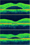
Methods: Five patients with chronic central serous chorioretinopathy were prospectively enrolled in this pilot study. Patients were administered finasteride (5 mg) daily for 3 months, after which study medication was withheld and patients were observed for 3 months. Main outcome measures included best-corrected visual acuity, central subfield macular thickness, and subretinal fluid volume as assessed by optical coherence tomography. Serum dihydrotestosterone, serum testosterone, and urinary cortisol were also measured.
Results: There was no change in mean best-corrected visual acuity. Mean center-subfield macular thickness and subretinal fluid volume reached a nadir at 3 months and rose to levels that were below baseline by 6 months. The changes in both optical coherence tomography parameters paralleled those in serum dihydrotestosterone level. In four patients, center-subfield macular thickness and/or subretinal fluid volume increased after discontinuation of finasteride. In the remaining patient, both optical coherence tomography parameters normalized with finasteride and remained stable when the study medication was discontinued.
Conclusion: Finasteride may represent a novel medical treatment for chronic central serous chorioretinopathy. Larger controlled clinical trials are needed to further assess the efficacy of finasteride for the treatment of central serous chorioretinopathy.
Figures


References
-
- Imamura Y, Fujiwara T, Margolis R, et al. Enhanced depth imaging optical coherence tomography of the choroid in central serous chorioretinopathy. Retina. 2009;29:1469–73. - PubMed
-
- Iida T, Kishi S, Hagimura N, et al. Persistent and bilateral choroidal vascular abnormalities in central serous chorioretinopathy. Retina. 1999;19:508–12. - PubMed
-
- Prunte C, Flammer J. Choroidal capillary and venous congestion in central serous chorioretinopathy. Am J Ophthalmol. 1996;121:26–34. - PubMed
-
- Giovannini A, Scassellati-Sforzolini B, D’Altobrando E, et al. Choroidal findings in the course of idiopathic serous pigment epithelium detachment detected by indocyanine green videoangiography. Retina. 1997;17:286–93. - PubMed
-
- von Ruckmann A, Fitzke FW, Fan J, et al. Abnormalities of fundus autofluorescence in central serous retinopathy. Am J Ophthalmol. 2002;133:780–6. - PubMed
MeSH terms
Substances
Grants and funding
LinkOut - more resources
Full Text Sources
Medical

