Review
. 2011 Jan 28;17(4):433-43.
doi: 10.3748/wjg.v17.i4.433.

Differentiation of Crohn's disease from intestinal tuberculosis in India in 2010

Affiliations
Review

Differentiation of Crohn's disease from intestinal tuberculosis in India in 2010

Anna Benjamin Pulimood et al. World J Gastroenterol. .

Abstract

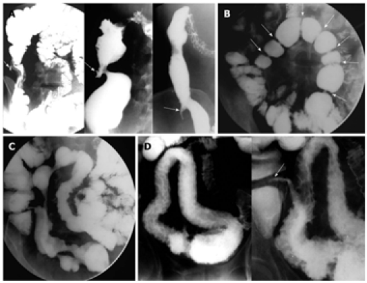
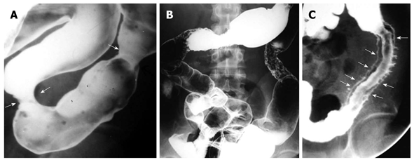
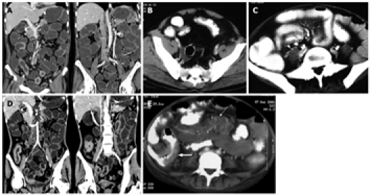
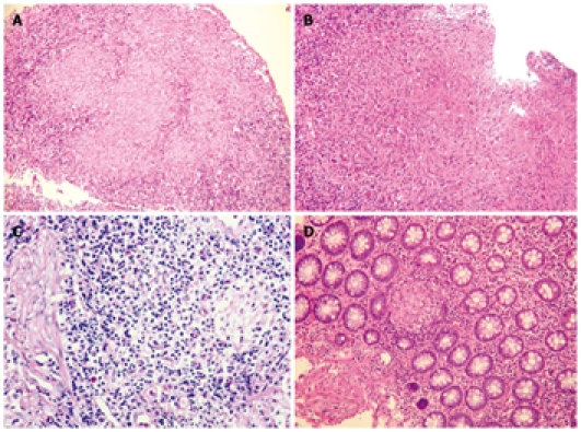
Differentiating intestinal tuberculosis from Crohn's disease (CD) is an important clinical challenge of considerable therapeutic significance. The problem is of greatest magnitude in countries where tuberculosis continues to be highly prevalent, and where the incidence of CD is increasing. The final clinical diagnosis is based on a combination of the clinical history with endoscopic studies, culture and polymerase chain reaction for Mycobacterium tuberculosis, biopsy pathology, radiological investigations and response to therapy. In a subset of patients, surgery is required and intraoperative findings with pathological study of the resected bowel provide a definitive diagnosis. Awareness of the parameters useful in distinguishing these two disorders in each of the different diagnostic modalities is crucial to accurate decision making. Newer techniques, such as capsule endoscopy, small bowel enteroscopy and immunological assays for Mycobacterium tuberculosis, have a role to play in the differentiation of intestinal tuberculosis and CD. This review presents currently available evidence regarding the usefulness and limitations of all these different modalities available for the evaluation of these two disorders.

Keywords: Clinical features; Crohn’s disease; Endoscopy; Enteroscopy; Histology; Radiology; Serology; Surgery; Therapy; Tuberculosis.


Figures

Figure 1
Figure 1
Findings on barium study of the small intestine. A: Barium study in three different patients with tuberculosis (TB) demonstrating shrunken, conical and retracted cecum (arrows); B: Multiple strictures (arrows) in ileal loops of a patient with TB. Note the short, concentric and smooth outline of the strictures; C: Multiple eccentric strictures seen in the ileum of a patient with Crohn's disease (CD). Note the normal ileo-cecal junction; D: Barium study showing an ulceronodular pattern involving a long segment of the ileum in a patient with CD. Note sparing of the ileo-cecal junction (arrow).
Figure 2
Figure 2
Findings on barium study of the colon in tuberculosis and Crohn's disease. A: Barium enema showing tubercular strictures (arrows) of the colon; B: Barium enema demonstrating skip lesions affecting descending and transverse colon as ulcers with areas of narrowing in a patient with Crohn's disease (CD); C: Deep ulcers with double tracking of sigmoid colon in a patient with CD (arrows).
Figure 3
Figure 3
Findings on computed tomography. A: Computed tomography (CT) enteroclysis with negative oral contrast showing mural thickening of ileum with skip areas and sparing of cecum in a patient with Crohn's disease (CD); B: Contrast-enhanced CT scan (CECT) showing asymmetrical mural thickening in ileal loop with deep ulcerations. Note fibro-fatty proliferation of mesentery; C: CECT in another patient with CD showing mesenteric vascular engorgement (Comb sign) with fibro-fatty proliferation of the mesentery; D: CT enteroclysis with negative oral contrast showing contiguous mural thickening of the terminal ileum and cecum in a patient with tuberculosis (TB); E: CECT in a patient with TB showing mural thickening of the terminal ileum and cecum (thick arrow) with multiple enlarged mesenteric lymph nodes showing central hypoattenuating and peripherally enhancing rims (thin arrow).
Figure 4
Figure 4
Histological features. A: Confluent granulomas in inflammatory granulation tissue from ulcerated colonic mucosa of a patient with tuberculosis (TB) [Hematoxylin and eosin (HE), 100 ×]; B: Large granuloma in the ulcerated mucosa of a patient with TB (HE, 100 ×); C: Microgranuloma composed of a small aggregate of macrophages in a lymphoid follicle from the mucosa of a patient with Crohn's disease (CD) (HE, 400 ×); D: Small pericryptal granuloma in the colonic mucosa of a patient with CD (HE, 100 ×).

References

    1. Pulimood AB, Peter S, Ramakrishna B, Chacko A, Jeyamani R, Jeyaseelan L, Kurian G. Segmental colonoscopic biopsies in the differentiation of ileocolic tuberculosis from Crohn's disease. J Gastroenterol Hepatol. 2005;20:688–696. - PubMed
    1. Lakatos PL. Recent trends in the epidemiology of inflammatory bowel diseases: up or down? World J Gastroenterol. 2006;12:6102–6108. - PMC - PubMed
    1. Pulimood AB, Ramakrishna BS, Kurian G, Peter S, Patra S, Mathan VI, Mathan MM. Endoscopic mucosal biopsies are useful in distinguishing granulomatous colitis due to Crohn's disease from tuberculosis. Gut. 1999;45:537–541. - PMC - PubMed
    1. Pai CG, Khandige GK. Is Crohn's disease rare in India? Indian J Gastroenterol. 2000;19:17–20. - PubMed
    1. Almadi MA, Ghosh S, Aljebreen AM. Differentiating intestinal tuberculosis from Crohn's disease: a diagnostic challenge. Am J Gastroenterol. 2009;104:1003–1012. - PubMed

MeSH terms