

Role of toll-like receptors and their downstream molecules in the development of nonalcoholic Fatty liver disease
- PMID: 21274430
- PMCID: PMC3026974
- DOI: 10.1155/2010/362847
Role of toll-like receptors and their downstream molecules in the development of nonalcoholic Fatty liver disease
Abstract
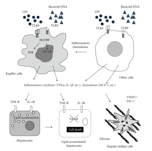
Activation of innate immunity is associated with the development of liver disease, including non-alcoholic fatty liver disease (NAFLD). In the innate immune system, Toll-like receptors (TLRs) are sensors that recognize bacterial and viral components such as lipopolysaccharide, bacterial DNA, and peptidoglycan. Recent data have demonstrated that the liver is exposed to a high load of TLR ligands due to bacterial overgrowth and increased intestinal permeability in NAFLD. Upon stimulation by these TLR ligands, hepatic immune cells produce various mediators that are involved in host defense. On the other hand, these mediators alter lipid metabolism, insulin signaling, and cell survival. Indeed, some TLR-deficient mice demonstrate lesser degrees of NAFLD even though TLR ligands are increased. This paper will highlight the recent progress on the study of TLR signaling and their downstream molecules in the development of NAFLD.
Figures
References
-
- Marra F, Gastaldelli A, Svegliati Baroni G, Tell G, Tiribelli C. Molecular basis and mechanisms of progression of non-alcoholic steatohepatitis. Trends in Molecular Medicine. 2008;14(2):72–81. - PubMed
-
- Ludwig J, Viggiano TR, McGill DB, Ott BJ. Nonalcoholic steatohepatitis. Mayo Clinic experiences with a hitherto unnamed disease. Mayo Clinic Proceedings. 1980;55(7):434–438. - PubMed
-
- Hilden M, Christoffersen P, Juhl E, Dalgaard JB. Liver histology in a ’normal’ population—examinations of 503 consecutive fatal traffic casualties. Scandinavian Journal of Gastroenterology. 1977;12(5):593–597. - PubMed
-
- Nomura H, Kashiwagi S, Hayashi J, Kajiyama W, Tani S, Goto M. Prevalence of fatty liver in a general population of Okinawa, Japan. Japanese Journal of Medicine. 1988;27(2):142–149. - PubMed
-
- Bellentani S, Saccoccio G, Masutti F, et al. Prevalence of and risk factors for hepatic steatosis in northern Italy. Annals of Internal Medicine. 2000;132(2):112–117. - PubMed
