. 2011:2011:510827.
doi: 10.1155/2011/510827. Epub 2011 Jan 10.

Loss of the NHE2 Na+/H+ exchanger in mice results in dilation of folliculo-stellate cell canaliculi

Affiliations

Loss of the NHE2 Na+/H+ exchanger in mice results in dilation of folliculo-stellate cell canaliculi

Marian L Miller et al. J Biomed Biotechnol. 2011.

Abstract

Genetic ablation of the NHE2 Na+/H+ exchanger causes gastric achlorhydria, absorptive defects in kidney and colon, and low fertility. Here we show that NHE2 is expressed in the pituitary, with the highest mRNA expression in pars distalis and lower expression in pars intermedia. In pars distalis of NHE2-null mice, prominent cyst-like dilatations of folliculo-stellate (FS) cell canaliculi developed with age, and there were increased FS cell area, accumulation of lipid in FS cell cytoplasm, redundancies in FS cell basement membrane, and other changes. The expansion of the canaliculi indicates that NHE2 is a major absorptive Na+/H+ exchanger in the luminal membranes lining the extensive network of channels formed by FS cells, which may provide a means of intrapituitary communication. The results suggest that NHE2 contributes to homeostatic regulation of the volume and composition of the canalicular fluid and may counter the secretory activity of the CFTR Cl⁻ channel, which is known to be expressed in pituitary.


Figures

Figure 1
Figure 1
Cystic changes develop with age in the pars distalis of Nhe2−/− mice. H&E stained sections of Nhe2−/− pituitaries were examined at the following ages: (a) 5 weeks, (b) 8 weeks, and (c) 10 weeks. Note the presence of dilated canaliculi (open spaces) at 8 weeks and 10 weeks. No cyst-like dilatations of the canaliculi were seen in the WT mice at 10 weeks (d) or earlier ages (not shown). Bar: 20 μm.
Figure 2
Figure 2
Northern blot analysis of NHE2 mRNA in pituitary and colon. Each lane contained 20 μg of total RNA from WT tissues. Hybridization with a rat NHE2 cDNA probe, which exhibited 93% identity to the mouse sequence and spanned the coding region of the gene, identified the expected 4.3 kb mRNA.
Figure 3
Figure 3
In situ hybridization analysis of NHE2 mRNA expression and histology. (a), (c) and (b), (d) are paired serial light micrographs of WT and Nhe2−/− pituitary, H&E-stained and in situ hybridization using the antisense probe (resp.). Hybridization with the sense probe yielded no specific signal in either WT or Nhe2−/− samples (data not shown). Three distinct areas of the pituitary are visible in both genotypes (white arrows) at 2x magnification. (e), (g) and (f), (h) are similar pairs at 10x showing that the most dense grain deposits were over the pars distalis (PD), less grain density over the pars intermedia (PI), and much lower grain density over the pars nervosa (PN). Black arrows in (e) and (g) indicate projections from the PI into the PN. The grain deposition over the PI demonstrated a distinct glandular (tubular) arrangement. (i) and (j) are paired WT and Nhe2−/− H&E stained sections at 40x where the difference in thickness of the PI (double headed white arrows) is clear. Cystic dilations (open spaces) of the FS cell canaliculi are clearly apparent in the PD of the Nhe2−/− mice ((b), (d), (f), (h), and (j)); bars : 750 μm in (a)–(d), 40 μm in (e)–(h), and 10 μm in (i) and (j).
Figure 4
Figure 4
Loss of NHE2 leads to increased Vd of both FS cells + cystic dilations (combined) and large granule cells. (a) and (b) are toluidine blue stained sections of pars distalis from WT and Nhe2−/− pituitaries. Arrows point to large granule cells (mainly somatotrophs and mammotrophs). Cystic dilations of FS cell canaliculi (C) developed with the loss of NHE2 and increased in size with age. Bar: 20 μm for both WT and Nhe2−/−. When morphometric data ((c), (d)) for Nhe2−/− (n = 9) and WT (n = 7) mice of all ages were considered, the Vd of the canalicular space plus FS cell nuclei and cytoplasm was significantly greater in Nhe2−/− than in WT; *P = .0079. The Vd of large-granule cells also increased significantly in Nhe2−/− mice; *P = .0026.
Figure 5
Figure 5
Nhe2−/− FS cells exhibited increased Vd but relatively normal ultrastructure. (a): morphometric data from the same animals and tissue sections used in Figure 4 showed a significant increase in the Vd of FS cells (cytoplasm + nucleus) in the Nhe2−/− pars distalis (*P = .013). Electron microscopy of WT and Nhe2−/− FS cells revealed normal ultrastructure except for an increase in lipid droplets (arrows) and decrease in desmosomal-mitochondrial associations (see Figures 6 and 9, resp.). FS cell junctional complexes (arrowheads) were numerous but seemingly unchanged by the loss of NHE2. Note the expanded canalicular space (C) in the Nhe2−/− micrograph; FS: folliculo-stellate cell; N: FS cell nucleus; arrows: lipid droplets; bar: 20 μm.
Figure 6
Figure 6
The Vd of lipid in FS and other cells in pars distalis was significantly increased. (a): morphometric data from the same animals and tissue sections used in Figure 4 showed a significant increase in the Vd of lipid droplets in Nhe2−/− mice when age groups were combined (*P = .0001), but was particularly striking in older Nhe2−/− mice. (b) and (c) are light micrographs, for which the bar is 20 μm; (d) is an electron micrograph, bar: 1 μm. Black arrows point to lipid droplets; C: canalicular space; N: nucleus.
Figure 7
Figure 7
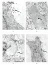
Microvilli on the apical membrane of FS cells of WT and Nhe2−/− mice were similar morphologically. Electron micrographs of (a) WT and (b)–(d) Nhe2−/− FS cells. Arrowheads point to microvilli. Dense patches of tightly packed microvilli were occasionally seen in FS cell canaliculi in the null animals; in (b), the arrowhead lies in the center of one such canaliculus. Cytoplasmic vacuoles in the apical cytoplasm of FS cells sometimes had membranous inclusions, as indicated by the arrow in (c). C: canaliculus; N: nucleus; bars: 1 μm.
Figure 8
Figure 8
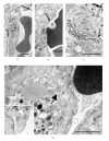
Duplicated and redundant basement membranes were found in Nhe2−/− pars distalis. In WT pars distalis (a) there are two parallel basement membranes between capillary endothelial cells and FS cells, one from each cell type. Similar parallel basement membranes were observed in some Nhe2−/− electron micrographs (b), but in conspicuous spots the basement membrane appeared to be duplicated and/or redundant in Nhe2−/− mice, which was particularly evident proximate to the FS cells ((c) and (d)), as indicated by the arrowhead in (d). Dotted lines overlay basement membranes. (a)–(c) black arrows point to fenestrated capillaries, which apparently did not change in Nhe2−/− mice. L: lipid droplet. Inset: white arrow, collagen. Bars: 1 μm.
Figure 9
Figure 9
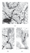
Nhe2−/− FS cells exhibited a reduction in the number of desmosomes associated with mitochondria. Desmosomal-mitochondrial associations can be seen in WT ((a) and (b)) and in Nhe2−/− mice (c). Desmosomes are often associated with one or two mitochondria (one from each cell). A significant decrease in desmosomal-mitochondrial associations was found in the FS cells of Nhe2−/− mice (see Table 1). Arrows point to mitochondria whose outer membranes are juxtaposed to intracellular filaments of desmosomes. Bars: 1 μm.

References

    1. Herbert DC, Silverman AY. Topographical distribution of the gonadotrophs, mammotrophs, somatotrophs and thyrotrophs in the pituitary gland of the baboon (Papio cynocephalus) Cell and Tissue Research. 1983;230(1):233–238. - PubMed
    1. Allaerts W, Carmeliet P, Denef C. New perspectives in the function of pituitary folliculo-stellate cells. Molecular and Cellular Endocrinology. 1990;71(2):73–81. - PubMed
    1. Inoue K, Couch EF, Takano K, Ogawa S. The structure and function of folliculo-stellate cells in the anterior pituitary gland. Archives of Histology and Cytology. 1999;62(3):205–218. - PubMed
    1. Ferrara N, Fujii DK, Goldsmith PC. Transport epithelial characteristics of cultured bovine pituitary follicular cells. American Journal of Physiology. 1987;252(3, part 1):E304–E312. - PubMed
    1. Ferrara N, Gospodarowicz D. Regulation of ion transport in hypophysial pars intermedia follicular cell monolayers. Biochemical and Biophysical Research Communications. 1988;157(3):1376–1382. - PubMed

Publication types

MeSH terms

LinkOut - more resources