. 2011 Jun;120(3):227-44.
doi: 10.1007/s00412-010-0306-5. Epub 2011 Jan 28.

Chromatin configuration and epigenetic landscape at the sex chromosome bivalent during equine spermatogenesis

Affiliations

Chromatin configuration and epigenetic landscape at the sex chromosome bivalent during equine spermatogenesis

Claudia Baumann et al. Chromosoma. 2011 Jun.

Abstract

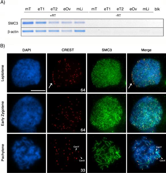
Pairing of the sex chromosomes during mammalian meiosis is characterized by the formation of a unique heterochromatin structure at the XY body. The mechanisms underlying the formation of this nuclear domain are reportedly highly conserved from marsupials to mammals. In this study, we demonstrate that in contrast to all eutherian species studied to date, partial synapsis of the heterologous sex chromosomes during pachytene stage in the horse is not associated with the formation of a typical macrochromatin domain at the XY body. While phosphorylated histone H2AX (γH2AX) and macroH2A1.2 are present as a diffuse signal over the entire macrochromatin domain in mouse pachytene spermatocytes, γH2AX, macroH2A1.2, and the cohesin subunit SMC3 are preferentially enriched at meiotic sex chromosome cores in equine spermatocytes. Moreover, although several histone modifications associated with this nuclear domain in the mouse such as H3K4me2 and ubH2A are conspicuously absent in the equine XY body, prominent RNA polymerase II foci persist at the sex chromosomes. Thus, the localization of key marker proteins and histone modifications associated with the XY body in the horse differs significantly from all other mammalian systems described. These results demonstrate that the epigenetic landscape and heterochromatinization of the equine XY body might be regulated by alternative mechanisms and that some features of XY body formation may be evolutionary divergent in the domestic horse. We propose equine spermatogenesis as a unique model system for the study of the regulatory networks leading to the epigenetic control of gene expression during XY body formation.


Figures

Fig. 1
Fig. 1
Nuclear morphology and cytological appearance of chromatin domains in mouse and horse spermatocyte nuclei indicate major differences in chromatin organization. a DAPI-stained pachytene spermatocyte from the mouse. DAPI-bright heterochromatin domains (asterisks) are clearly distinguishable from DAPI-pale euchromatic areas. c The position of the sex chromosome-containing XY body is distinctly visible by DAPI staining (arrows in a and c) and co-localizes with the signal obtained following fluorescence in situ hybridization (FISH) using an X chromosome-specific probe (red). b Typical chromatin organization in pachytene spermatocytes from the horse. Heterochromatin and euchromatin domains are indistinguishable by DAPI staining. d Similarly, the localization of the XY body (arrow) is not discernible by DAPI staining and requires co-localization with the signal obtained by FISH using a horse-specific X chromosome probe (red). Arrowheads indicate the position of synapsed bivalents following staining with SMC3 (green) during pachytene stage. Scale bar=10 μm
Fig. 2
Fig. 2
Analysis of meiotic configuration in equine spermatocytes. a Expression of the structural maintenance of chromosomes 3 (SMC3) coding sequences in mouse (mT) and horse (eT) testis tissue, equine fetal ovary (eOv), and mouse liver tissue (mLi) by RT-PCR. β-actin mRNA expression was used as housekeeping control. No amplification was detectable under omission of reverse transcriptase (−RT) or in the negative control (blk). b Immunochemical localization of kinetochore proteins (CREST, red) and synaptonemal complex components (SMC3, green) in equine spermatocytes reveals the position of the centromere in each of the 64 asynapsed equine chromosomes during leptotene. Note the arrangement of telocentric chromosome bivalents on a bouquet configuration (arrow, upper panel). At the zygotene stage (middle panel), centromeres become redistributed throughout the nucleoplasm. The number of CREST signals decreases to 33 at the pachytene stage (lower panel), indicating complete synapsis between 31 autosomal chromosome pairs. Pairing of the sex chromosomes, however, remains restricted to a distal pseudoautosomal region and centromeres remain separated as indicated by two smaller CREST foci (CenY and CenX) (lower panel, arrowheads). DNA is shown in blue. Scale bar=10 μm
Fig. 3
Fig. 3
The cohesin subunit SMC3 is enriched at the sex chromosome bivalent in equine pachytene spermatocytes. a SMC3 (green) marks cohesin axes at synapsed chromosomes (arrowheads) in mouse and horse spermatocytes. In horse nuclei, SMC3 is preferentially enriched at the sex chromosome bivalent (arrow and inset, lower panel) as evidenced by co-localization with the signal originating from an X chromosome-specific probe (red). DNA is shown in blue. Scale bar= 10μm. b Ratio of fluorescence intensities at sex chromosome bivalents (n=10 for each species) compared to autosomes in equine and mouse spermatocytes, respectively
Fig. 4
Fig. 4
Association of γH2AX with meiotic sex chromosome cores in equine pachytene spermatocytes. a During leptotene (upper panel) and zygotene (middle panel) stages of meiosis, γH2AX (red) exhibits a diffuse nucleoplasmic staining indicating the presence of DNA double strand breaks. At the pachytene stage, γH2AX is found associated primarily with sex chromosome cores (lower panel, arrow). b In contrast to mouse pachytene spermatocytes, which exhibit a prominent, cloud-like, association of γH2AX with the macrochromatin domain of the XY body (left image, arrow), horse pachytene spermatocytes exhibit γH2AX staining at the sex chromosome cores only (right image, arrow). c Determination of Mander's co-localization coefficients (R) to quantify the levels of γH2AX staining (red) beyond sex chromosome cores (green) in mouse and horse pachytene spermatocytes. d Percentage of signal overlap between γH2AX and SMC3 at mouse and horse XY bodies (n=10 for each species), demonstrating that γH2AX staining extends beyond meiotic chromosome cores and occupies the entire macrochromatin domain in the mouse. In striking contrast, a higher degree of co-localization indicates that γH2AX is essentially restricted to XY chromosome cores in horse pachytene spermatocytes. DNA is shown in blue and SMC3 in green. Scale bar=10 μm
Fig. 5
Fig. 5
Epigenetic modifications at the sex chromosome bivalent in equine pachytene spermatocytes. a Histone H3 dimethylation at lysine 4 (H3K4me2; green) exhibits a diffuse nucleoplasmic staining but is excluded from pericentric heterochromatin domains in mouse pachytene spermatocytes (arrowhead). Note the prominent accumulation of H3K4me2 at the macrochromatin domain and its co-localization with γH2AX (red) in mouse nuclei (arrow). In contrast, the equine XY body is completely devoid of this histone modification (arrow, lower panel), while the localization to euchromatic domains is conserved. b The mono-ubiquitinated form of histone H2A (ubH2A, red) co-localizes with telomeric domains (arrowheads, insets) in mouse (upper panel) and horse (lower panel) pachytene spermatocytes and shows a prominent accumulation at the XY body in the mouse (arrow). In the horse, ubH2A exhibits a diffuse nucleoplasmic staining with no preferential enrichment at sex chromatin (arrow, lower panel). c The histone variant macroH2A1.2 (green) is co-localized with γH2AX (red) at the macrochromatin domain and shows a preferential enrichment at DAPI-bright pericentric heterochromatin (arrowhead) in mouse spermatocytes. In equine spermatocytes, macroH2A1.2 labels lateral elements of the synaptonemal complex in all chromosome bivalents and is exclusively associated chromosome cores at the XY body (arrow). DNA is shown in blue. Scale bars=10 μm
Fig. 6
Fig. 6
Persistence of RNA polymerase II at the XY body in equine spermatocytes. a The pre-messenger RNA splicing factor Smith antigen (red) is present in the nucleoplasm of mouse (upper panel) and horse (lower panel) spermatocytes, but is excluded from the XY body in both species (arrow). b RNA polymerase II (red) exhibits a diffuse nucleoplasmic staining in mouse (arrowhead, upper panel) and horse (lower panel) pachytene spermatocytes. In mouse spermatocytes, RNA polymerase II is excluded from the XY body (arrow) in the majority of cells, which is consistent with global transcriptional silencing of this chromatin domain. In contrast, RNA polymerase II foci (arrowhead) persist associated with sex chromosome cores in equine spermatocytes c Proportion of equine spermatocytes with RNA polymerase II staining. DNA is shown in blue. Scale bar=10 μm
Fig. 7
Fig. 7
Detection of meiotic recombination events in equine sperma-▶tocytes. a Simultaneous staining of CREST (red) and SMC3 (green) reveals the presence of fully synapsed acrocentric and submetacentric bivalents at the pachytene stage. Note that centromeric domains are distributed throughout the nucleoplasm. The pseudoautosomal region (PAR) is established between the distant segments of an acrocentric Y chromosome (dark green in diagram) and the submetacentric X chromosome (light green). The position of the sex chromosome centromeres (CenY and CenX) is indicated. b Rad51 foci (red) are detected in early pachytene spermatocytes. Each bivalent exhibits one to two Rad51 foci along the chromosome axes (bold arrow). The sex chromosomes (arrow) exhibit a single Rad51 focus at the PAR (arrowhead). c The mismatch repair protein MLH1 (red) marks the sites of crossover formation along chromosome cores of all autosomes (bold arrow). Moreover, the partially synapsed sex chromosomes (arrow) show a single MLH1 focus in the majority of cells (arrowhead). Scale bar=10 μm. d Scatter plot diagram to visualize the average numbers of MLH foci per chromosome bivalent (2n=64) detected in equine pachytene spermatocytes (n=33). DNA is shown in blue. Scale bar=10 μm
Fig. 8
Fig. 8
Comparison of epigenetic modifications at the XY body in mouse and equine pachytene spermatocytes. The chromatin domain at the XY body in equine pachytene spermatocytes demonstrates evolutionary divergence in both, morphological appearance and in regard to its epigenetic landscape. Solid arrows indicate enrichment; broken lines designate absence of enrichment or exclusion from the XY body domain

Similar articles

Cited by

References

    1. Akhmedov A, Gross B, Jessberger R. Mammalian smc3 c-terminal and coiled-coil protein domains specifically bind palindromic DNA, do not block DNA ends, and prevent DNA bending. J Biol Chem. 1999;274(53):38216–38224. - PubMed
    1. Anderson SF, Schlegel BP, Nakajima T, Wolpin ES, Parvin JD. BRCA1 protein is linked to the RNA polymerase Π holoenzyme complex via RNA helicase A. Nat Genet. 1998;19:254–256. - PubMed
    1. Baarends W, Hoogerbrugge J, Roest H, Ooms M, Vreeburg J, Hoeijmakers J, Grootegoed J. Histone ubiquitination and chromatin remodeling in mouse spermatogenesis. Dev Biol. 1999;207(2):322–333. - PubMed
    1. Baarends W, Wassenaar E, van der Laan R, Hoogerbrugge J, Sleddens-Linkels E, Hoeijmakers J, de Boer P, Grootegoed J. Silencing of unpaired chromatin and histone H2A ubiquitination in mammalian meiosis. Mol Cell Biol. 2005;25(3):1041–1053. - PMC - PubMed
    1. Bannister L, Schimenti J. Homologous recombinational repair proteins in mouse meiosis. Cytogenet Genome Res. 2004;107(3–4):191–200. - PubMed

Publication types