

Relationship between cytomegalovirus (CMV) IgG serology, detectable CMV DNA in peripheral monocytes, and CMV pp65(495-503)-specific CD8+ T cells in older adults
- PMID: 21274637
- PMCID: PMC3220402
- DOI: 10.1007/s11357-011-9205-9
Relationship between cytomegalovirus (CMV) IgG serology, detectable CMV DNA in peripheral monocytes, and CMV pp65(495-503)-specific CD8+ T cells in older adults
Abstract
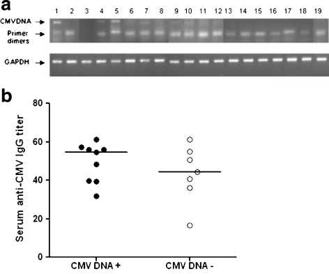
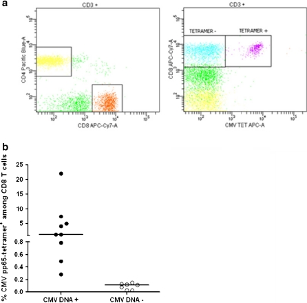
In immunocompetent individuals, cytomegalovirus (CMV) is thought to persist in a latent state in monocytes and myeloid progenitor cells, establishing a lifelong infection. In CMV-seropositive older adults, aging has been associated with both expansion of CMV pp65(495-503)-specific CD8(+) T cell clones and shrinkage of the T cell repertoire that characterize T cell immunosenescence. In fact it has been suggested that chronic CMV infection is a driving force in age-related T cell immunosenescence. In older adults, chronic CMV infection is conventionally diagnosed by positive IgG serology which does not distinguish between past and persistent infections. To better define the relationship between chronic CMV infection and expansion of CMV pp65(495-503)-specific CD8(+) T cells, we directly assessed CMV viral DNA in monocyte-enriched peripheral blood mononuclear cells in 16 HLA-A2-positive elderly volunteers (mean age = 83 years). While all participants had positive CMV IgG serology by enzyme-linked immunosorbent assays, only nine (56%) had detectable CMV DNA by nested polymerase chain reaction. These nine individuals had significantly higher percentages of CMV pp65(495-503) tetramer-positive CD8(+) T cells (median = 1.3%) than those without detectable CMV DNA (median = 0.1%; p < 0.001). Absolute CMV IgG antibody titers did not differ between these two groups (median = 54.6 vs 44.2 EU/ml, respectively, p = 0.4). CMV IgM titers were negative for all 16 participants, suggesting that recent primary CMV infection was unlikely. These results demonstrate a strong association between the presence of CMV DNA in peripheral monocytes and the expansion of CD8(+) T cells specific for the CMV immunodominant epitope pp65(495-503). Although the sample size in this study is relatively small, these findings provide initial evidence suggesting the heterogeneity of CMV IgG-seropositive older adult population and CMV viral DNA detection in peripheral monocytes as an informative tool to better understand the relationship between chronic CMV infection and T cell immunosenescence.
Figures
References
-
- Bolovan-Fritts CA, Mocarski ES, Wiedeman JA. Peripheral blood CD14(+) cells from healthy subjects carry a circular conformation of latent cytomegalovirus genome. Blood. 1999;93:394–398. - PubMed
-
- Brooimans RA, Boyce CS, Popma J, Broyles DA, Gratama JW, Southwick PC, Keeney M. Analytical performance of a standardized single-platform MHC tetramer assay for the identification and enumeration of CMV-specific CD8+ T lymphocytes. Cytom A. 2008;73:992–1000. doi: 10.1002/cyto.a.20641. - DOI - PubMed
-
- Hadrup SR, Strindhall J, Kollgaard T, Seremet T, Johansson B, Pawelec G, thor Straten P, Wikby A. Longitudinal studies of clonally expanded CD8 T cells reveal a repertoire shrinkage predicting mortality and an increased number of dysfunctional cytomegalovirus-specific T cells in the very elderly. J Immunol. 2006;176:2645–2653. - PubMed
Publication types
MeSH terms
Substances
Grants and funding
LinkOut - more resources
Full Text Sources
Molecular Biology Databases
Research Materials
Miscellaneous