. 2011 Feb;53(2):628-39.
doi: 10.1002/hep.24041. Epub 2011 Jan 3.

Activation of alpha(1) -adrenergic receptors stimulate the growth of small mouse cholangiocytes via calcium-dependent activation of nuclear factor of activated T cells 2 and specificity protein 1

Affiliations

Activation of alpha(1) -adrenergic receptors stimulate the growth of small mouse cholangiocytes via calcium-dependent activation of nuclear factor of activated T cells 2 and specificity protein 1

Gianfranco Alpini et al. Hepatology. 2011 Feb.

Abstract

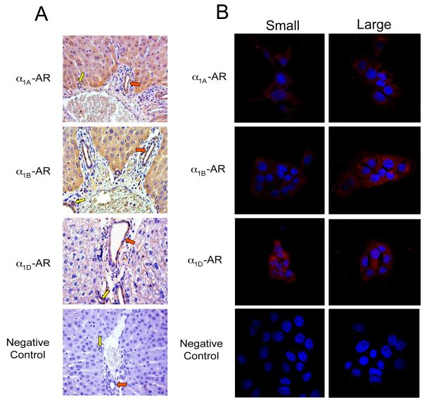
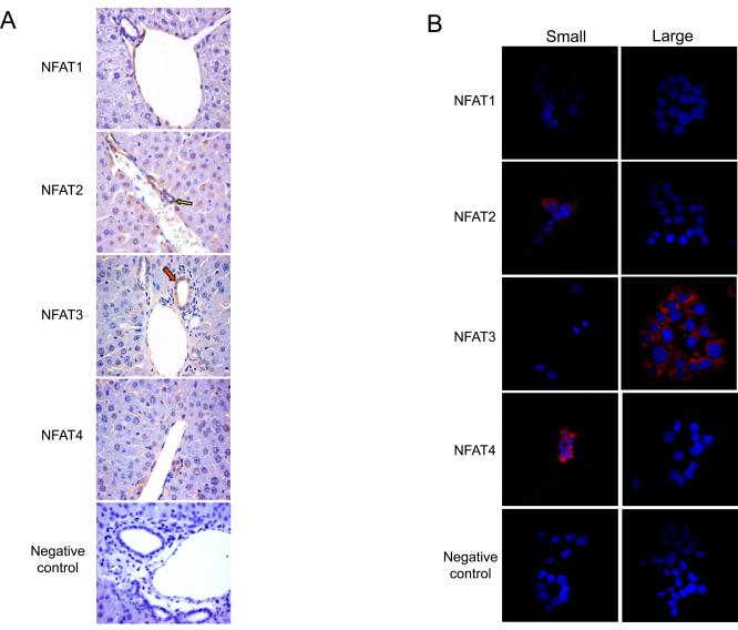
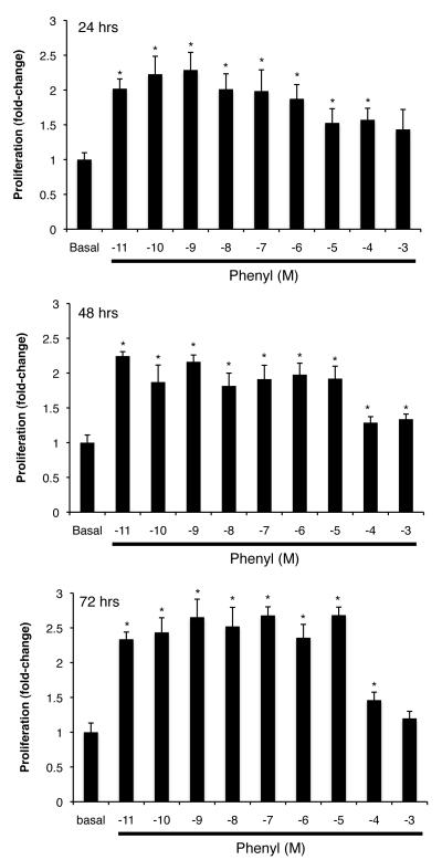
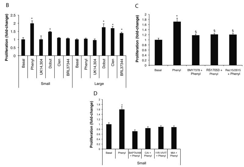
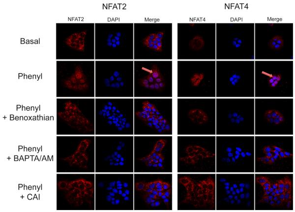
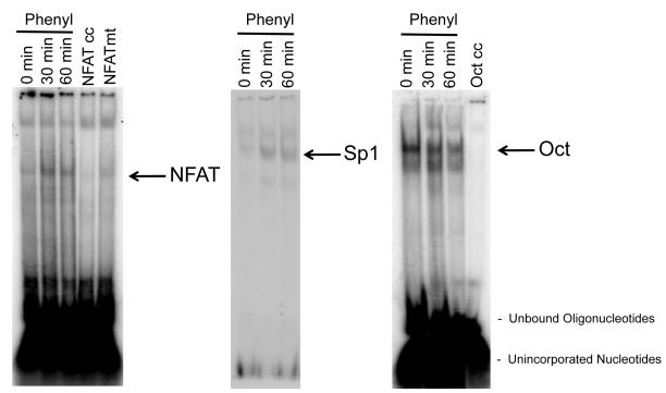
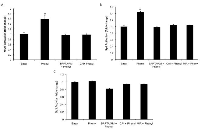
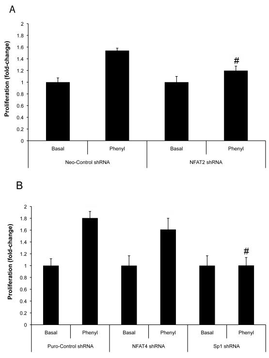
Small cholangiocytes proliferate via activation of calcium (Ca(2+) )-dependent signaling in response to pathological conditions that trigger the damage of large cyclic adenosine monophosphate-dependent cholangiocytes. Although our previous studies suggest that small cholangiocyte proliferation is regulated by the activation of Ca(2+) -dependent signaling, the intracellular mechanisms regulating small cholangiocyte proliferation are undefined. Therefore, we sought to address the role and mechanisms of action by which phenylephrine, an α(1) -adrenergic agonist stimulating intracellular D-myo-inositol-1,4,5-triphosphate (IP(3) )/Ca(2+) levels, regulates small cholangiocyte proliferation. Small and large bile ducts and cholangiocytes expressed all AR receptor subtypes. Small (but not large) cholangiocytes respond to phenylephrine with increased proliferation via the activation of IP(3) /Ca(2+) -dependent signaling. Phenylephrine stimulated the production of intracellular IP(3) . The Ca(2+) -dependent transcription factors, nuclear factor of activated T cells 2 (NFAT2) and NFAT4, were predominantly expressed by small bile ducts and small cholangiocytes. Phenylephrine stimulated the Ca(2+) -dependent DNA-binding activities of NFAT2, NFAT4, and Sp1 (but not Sp3) and the nuclear translocation of NFAT2 and NFAT4 in small cholangiocytes. To determine the relative roles of NFAT2, NFAT4, or Sp1, we knocked down the expression of these transcription factors with small hairpin RNA. We observed an inhibition of phenylephrine-induced proliferation in small cholangiocytes lacking the expression of NFAT2 or Sp1. Phenylephrine stimulated small cholangiocyte proliferation is regulated by Ca(2+) -dependent activation of NFAT2 and Sp1.

Conclusion: Selective stimulation of Ca(2+) -dependent small cholangiocyte proliferation may be key to promote the repopulation of the biliary epithelium when large bile ducts are damaged during cholestasis or by toxins.


Figures

Fig. 1
Fig. 1
Expression of α1A, α1B, α1D AR by immunohistochemistry (A) in liver sections, and immunofluorescence (B) in immortalized small and large cholangiocytes. (A) Small (yellow arrows) and large (red arrows) bile ducts were positive for α1A-, α1B-, and α1D-AR expression. Original magnification, x20. (B) Immortalized small and large cholangiocytes were positive for α1A-, α1B-, and α1D-AR. AR expression is shown in red with nuclei counterstained with DAPI (blue). Original magnification, x60. The negative control performed without the primary antibody is presented at the bottom of the figure. Bile ducts (A) and immortalized small and large cholangiocytes (B) express α1A, α1B, and α1D-AR.
Fig. 2
Fig. 2
Expression of NFAT isoforms by immunohistochemistry (A) in liver sections, and immunofluorescence (B) in immortalized small and large cholangiocytes. (A) NFAT2 and 4 were expressed predominantly by small bile ducts (yellow arrows; for semiquantitative data see Suppl. Table 1). A small % of cholangiocytes in large bile ducts (red arrows) stained positively for NFAT2 and 4. NFAT3 was expressed only in large bile ducts, whereas NFAT1 was not expressed in either sized bile ducts. Original magnification, x20. (B) A similar expression profile was observed in immortalized small and large cholangiocytes. Small cholangiocytes were positive for NFAT2 and 4. Large cholangiocytes were positive for NFAT3, whereas neither cell type expressed NFAT1. Original magnification, x60. A representative negative control performed without the primary antibody is presented at the bottom of the figure.
Fig. 3
Fig. 3
Expression of CK-19 by immunohistochemistry in liver sections of normal mouse treated with vehicle or phenylephrine for 1 week in the absence or presence of 11R-VIVIT or MiA. Large (red arrow) and small (yellow arrows) ducts are indicated. Original Magnification X 40. Measurement of IBDM of small and large cholangiocytes in liver sections from the selected groups of mice. Chronic administration of phenylephrine to normal mice induces a significant increase in IBDM of small but not large cholangiocytes, increase that was blocked by 11R-VIVIT and MiA. *p=0.0022 (by Mann-Whitney test) vs. IBDM of small cholangiocytes from normal mice treated with phenylephrine vs. normal mice treated with vehicle.
Fig. 4
Fig. 4
(A) Effect of different doses (10−11 to 10−3 M) of phenylephrine on the proliferation of immortalized small cholangiocytes. The doses (10−11 to 10−5 M) used for phenylephrine induced a similar increase in small cholangiocyte proliferation. *denotes significance (p<0.05) when compared with the respective basal treatment using a Kruskal-Wallis test (n=6-14). (B) In addition to phenylephrine, dobutamine increased small cholangiocyte proliferation. In immortalized large cholangiocytes, dobutamine, clenbuterol and BDL 37344 induced a significant increase in proliferation, whereas phenylephrine and UK14,304 had no effect. *denotes significance (p<0.05) when compared with the respective basal treatment using a Kruskal-Wallis test (n=14). (C) Effect of phenylephrine (10 μM for 24 hr) on the proliferation of immortalized small cholangiocytes in the absence or presence of selective AR antagonists. α1A-, α1B-, and α1D-AR antagonists induced a partial yet significant reduction in phenylephrine-induced small cholangiocyte proliferation. *denotes significance (p<0.05) when compared with the respective basal treatment using a t test (n=14). §denotes significance (p<0.05) when compared with phenylephrine-induced proliferation. (D) Phenylephrine stimulates small cholangiocytes proliferation in a Ca2+-dependent mechanism. Small mouse cholangiocytes were stimulated with phenylephrine in the presence/absence of BAPTA/AM; CAI; 11R-VIVIT; or MiA. Phenylephrine-stimulated small cholangiocyte proliferation was prevented by BAPTA/AM, CAI, 11R-VIVIT, or MiA. *denotes significance (p<0.05) when compared with the respective basal treatment using a Kruskal-Wallis test (n=14).
Fig. 4
Fig. 4
(A) Effect of different doses (10−11 to 10−3 M) of phenylephrine on the proliferation of immortalized small cholangiocytes. The doses (10−11 to 10−5 M) used for phenylephrine induced a similar increase in small cholangiocyte proliferation. *denotes significance (p<0.05) when compared with the respective basal treatment using a Kruskal-Wallis test (n=6-14). (B) In addition to phenylephrine, dobutamine increased small cholangiocyte proliferation. In immortalized large cholangiocytes, dobutamine, clenbuterol and BDL 37344 induced a significant increase in proliferation, whereas phenylephrine and UK14,304 had no effect. *denotes significance (p<0.05) when compared with the respective basal treatment using a Kruskal-Wallis test (n=14). (C) Effect of phenylephrine (10 μM for 24 hr) on the proliferation of immortalized small cholangiocytes in the absence or presence of selective AR antagonists. α1A-, α1B-, and α1D-AR antagonists induced a partial yet significant reduction in phenylephrine-induced small cholangiocyte proliferation. *denotes significance (p<0.05) when compared with the respective basal treatment using a t test (n=14). §denotes significance (p<0.05) when compared with phenylephrine-induced proliferation. (D) Phenylephrine stimulates small cholangiocytes proliferation in a Ca2+-dependent mechanism. Small mouse cholangiocytes were stimulated with phenylephrine in the presence/absence of BAPTA/AM; CAI; 11R-VIVIT; or MiA. Phenylephrine-stimulated small cholangiocyte proliferation was prevented by BAPTA/AM, CAI, 11R-VIVIT, or MiA. *denotes significance (p<0.05) when compared with the respective basal treatment using a Kruskal-Wallis test (n=14).
Fig. 5
Fig. 5
Phenylephrine stimulates the nuclear translocation of NFAT2 and NFAT4. Immortalized small cholangiocytes were stimulated with phenylephrine in the presence/absence of benoxathian, BAPTA/AM, and CAI for 60 minutes. By immunofluorescence, phenylephrine stimulates the nuclear translocation of NFAT2 and NFAT4 (arrows), which was blocked by benoxathian, BAPTA/AM, and CAI. Original magnification, x60.
Fig. 6
Fig. 6
Evaluation of phenylephrine-induced NFAT and Sp1 DNA-binding activity by EMSA. Immortalized small cholangiocytes were stimulated with phenylephrine for 0, 30 and 60 min at 37°C and DNA-binding activity was assessed by EMSA. Phenylephrine stimulated a time-dependent increase in DNA-binding for both NFAT (NFAT2 and 4 can both bind the consensus sequence) and Sp1. DNA-binding activity to the Oct consensus sequence was used as a loading control. Specificity of binding was demonstrated by adding 50 fold excess of either unlabeled NFAT consensus sequence, (NFAT cc), mutated NFAT consensus sequence (NFAT mt) or Oct sequence to the nuclear extract taken at time 0. NFAT = nuclear factor of activated T-cells; NFAT cc = NFAT cold consensus sequence; NFAT mt = NFAT mutated competitor; Oct = octamer binding factor; and Oct cc = Oct cold consensus sequence.
Fig. 7
Fig. 7
Phenylephrine induces NFAT2 and Sp1 (but not Sp3) DNA-binding activity. (A) Immortalized small cholangiocytes were stimulated with phenylephrine in the presence/absence of BAPTA/AM and CAI for 60 min. NFAT2 DNA-binding activity was determined by ELISA. Phenylephrine stimulates the DNA-binding activity of NFAT2, which is blocked by BAPTA/AM and CAI. *denotes significance (p<0.05) when compared with the respective basal treatment using a t test (n=4). (B-C) Small cholangiocytes were stimulated with phenylephrine in the presence/absence of BAPTA/AM, CAI and MiA for 60 min. Sp1 and Sp3 DNA-binding ac8ivity was determined by ELISA. Phenylephrine stimulates the DNA-binding activity of Sp1 (B) (but not Sp3 (C)), which is blocked by BAPTA/AM, CAI and MiA. *denotes significance (p<0.05) when compared with the respective basal treatment using a t test (n=4).
Fig. 8
Fig. 8
Knockdown of NFAT2 and Sp1 expression prevents phenylephrine-induced proliferation of immortalized small cholangiocytes. The expression of NFAT2, NFAT4, or Sp1 was knockdown in small cholangiocytes by stable transfection of the respective shRNA. The knockdown of NFAT2 (A) and Sp1 (B) resulted in the prevention of phenylephrine-stimulated small cholangiocyte proliferation. (B) Knockdown of the expression NFAT4 did not significantly inhibit phenylephrine stimulation of small cholangiocyte proliferation. Data was expressed as fold-change relative to the respective basal values. #denotes significance (p<0.05) when compared with the respective phenyl-stimulated treatment group using a t test (n=7).

References

    1. Alpini G, Prall RT, LaRusso NF. The pathobiology of biliary epithelia. In: Arias IM, Boyer JL, Chisari FV, Fausto N, Jakoby W, Schachter D, Shafritz DA, editors. The Liver; Biology & Pathobiology. 4th Ed Lippincott Williams & Wilkins; Philadelphia, PA: 2001. pp. 421–435.
    1. Alpini G, Glaser S, Ueno Y, Pham L, Podila PV, Caligiuri A, LeSage G, et al. Heterogeneity of the proliferative capacity of rat cholangiocytes after bile duct ligation. Am J Physiol Gastrointest Liver Physiol. 1998;274:G767–775. - PubMed
    1. LeSage G, Glaser S, Marucci L, Benedetti A, Phinizy JL, Rodgers R, Caligiuri A, et al. Acute carbon tetrachloride feeding induces damage of large but not small cholangiocytes from BDL rat liver. Am J Physiol Gastrointest Liver Physiol. 1999;276:G1289–1301. - PubMed
    1. Francis H, Glaser S, Ueno Y, LeSage G, Marucci L, Benedetti A, Taffetani S, et al. cAMP stimulates the secretory and proliferative capacity of the rat intrahepatic biliary epithelium through changes in the PKA/Src/MEK/ERK1/2 pathway. J Hepatol. 2004;41:528–537. - PubMed
    1. Ueno Y, Alpini G, Yahagi K, Kanno N, Moritoki Y, Fukushima K, Glaser S, et al. Evaluation of differential gene expression by microarray analysis in small and large cholangiocytes isolated from normal mice. Liver Int. 2003;23:449–459. - PubMed

Publication types

MeSH terms