. 2011 Feb;53(2):649-60.
doi: 10.1002/hep.24059. Epub 2011 Jan 10.

Interferon regulatory factor 3 and type I interferons are protective in alcoholic liver injury in mice by way of crosstalk of parenchymal and myeloid cells

Affiliations

Interferon regulatory factor 3 and type I interferons are protective in alcoholic liver injury in mice by way of crosstalk of parenchymal and myeloid cells

Jan Petrasek et al. Hepatology. 2011 Feb.

Abstract

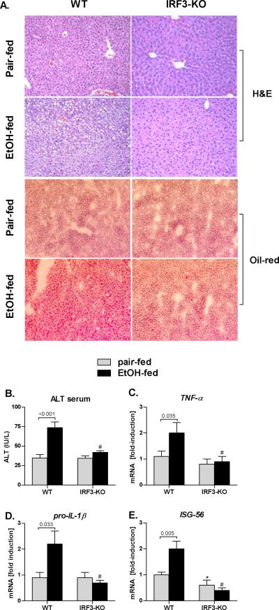
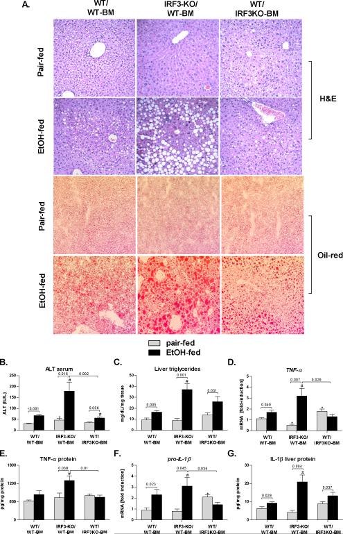
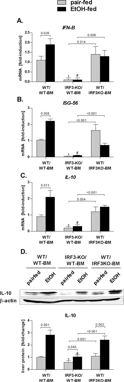
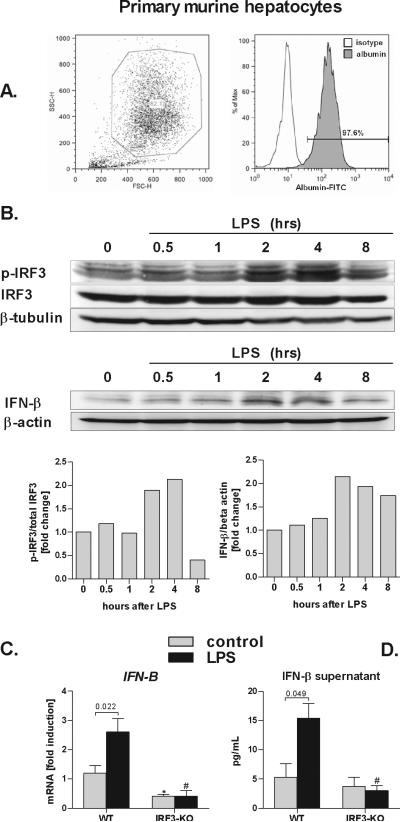
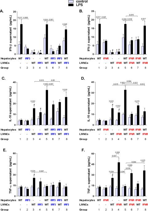
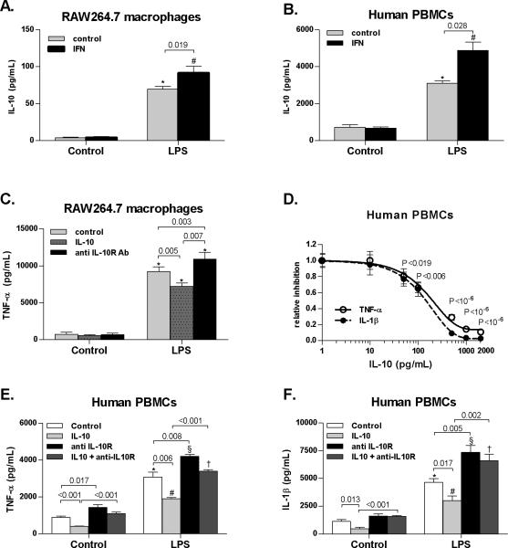
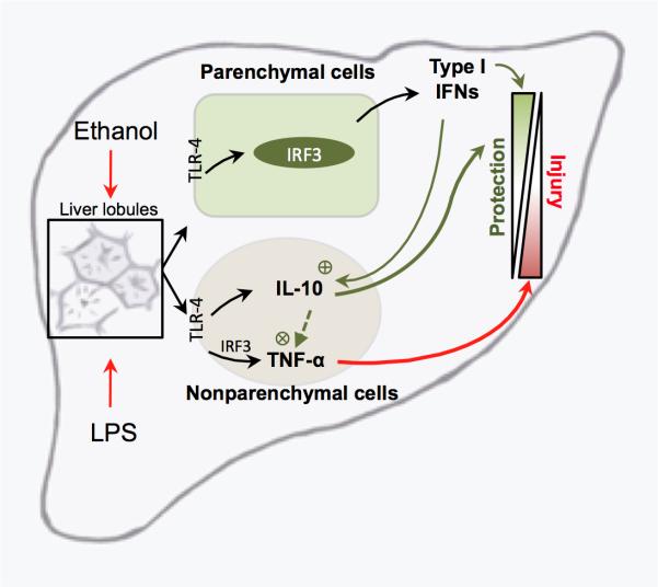
Alcoholic liver disease (ALD) features increased hepatic exposure to bacterial lipopolysaccharide (LPS). Toll-like receptor-4 (TLR4) recognizes LPS and activates signaling pathways depending on MyD88 or TRIF adaptors. We previously showed that MyD88 is dispensable in ALD. TLR4 induces Type I interferons (IFNs) in an MyD88-independent manner that involves interferon regulatory factor-3 (IRF3). We fed alcohol or control diets to wild-type (WT) and IRF3 knock-out (KO) mice, and to mice with selective IRF3 deficiency in liver parenchymal and bone marrow-derived cells. Whole-body IRF3-KO mice were protected from alcohol-induced liver injury, steatosis, and inflammation. In contrast to WT or bone marrow-specific IRF3-KO mice, deficiency of IRF3 only in parenchymal cells aggravated alcohol-induced liver injury, associated with increased proinflammatory cytokines, lower antiinflammatory cytokine interleukin 10 (IL-10), and lower Type I IFNs compared to WT mice. Coculture of WT primary murine hepatocytes with liver mononuclear cells (LMNC) resulted in higher LPS-induced IL-10 and IFN-β, and lower tumor necrosis factor alpha (TNF-α) levels compared to LMNC alone. Type I IFN was important because cocultures of hepatocytes with LMNC from Type I IFN receptor KO mice showed attenuated IL-10 levels compared to control cocultures from WT mice. We further identified that Type I IFNs potentiated LPS-induced IL-10 and inhibited inflammatory cytokine production in both murine macrophages and human leukocytes, indicating preserved cross-species effects. These findings suggest that liver parenchymal cells are the dominant source of Type I IFN in a TLR4/IRF3-dependent manner. Further, parenchymal cell-derived Type I IFNs increase antiinflammatory and suppress proinflammatory cytokines production by LMNC in paracrine manner.

Conclusion: Our results indicate that IRF3 activation in parenchymal cells and resulting type I IFNs have protective effects in ALD by way of modulation of inflammatory functions in macrophages. These results suggest potential therapeutic targets in ALD.


Figures

Fig. 1
Fig. 1. IRF3-deficiency protects against alcohol-induced liver damage
Wild-type (WT) and IRF3-deficient (IRF3-KO) were fed Lieber DeCarli ethanol or control (pair-fed) diet and sacrificed after 4 weeks. Livers were fixed in formalin and stained with H&E or with Oil-red-O; magnification 200x (A). Serum ALT levels (B) were analyzed. Messenger RNA levels of liver (C) tumor necrosis factor α (TNFA), (D) pro-interleukin 1-beta (IL-1β) and (E) interferon stimulated gene ISG56 were analyzed by real-time PCR and normalized to 18s.Values are shown as mean ± SEM fold increase over WT pair-fed control group (9-12 mice per group). Numbers in graphs denote p values; *) p < 0.05 vs. pair-fed WT mice; #) p < 0.05 vs. ethanol-fed WT mice.
Fig. 2
Fig. 2. Parenchymal cell-specific IRF3 deficiency aggravates alcohol-induced liver damage
Wild-type mice with transplanted WT bone marrow (WT/WT-BM), IRF3-deficient mice with transplanted wild-type bone marrow (IRF3-KO/WT-BM) and WT mice with transplanted IRF3-deficient bone marrow (WT/IRF3-KO) were fed Lieber DeCarli ethanol or control (pair-fed) diet and sacrificed after 4 weeks. Livers were fixed in formalin and stained with H&E or with Oilred-O; magnification 200x (A). Serum ALT levels (B) and liver triglycerides (C) were analyzed. Messenger RNA levels of liver (D) tumor necrosis factor α (TNFA) and (F) pro-interleukin 1-beta (IL-1β) were analyzed by real-time PCR and normalized to 18s. Liver TNF-α and IL-1β levels were analyzed using ELISA (E,G). Values are shown as mean ± SEM fold increase over wild-type pair-fed control group (11-17 mice per group). Numbers in graphs denote p values; *) p < 0.05 vs. pair-fed wild-type mice; #) p < 0.05 vs. ethanol-fed wild-type mice.
Fig. 3
Fig. 3. Parenchymal cell-specific IRF3 deficiency is associated with decreased Type I IFN and IL-10 induction
Wild-type mice with transplanted WT bone marrow (WT/WT-BM), IRF3-deficient mice with transplanted wild-type bone marrow (IRF3-KO/WT-BM) and WT mice with transplanted IRF3-deficient bone marrow (WT/IRF3-KO) were fed Lieber DeCarli ethanol or control (pair-fed) diet and sacrificed after 4 weeks. Messenger RNA levels of (A) liver interferon β (IFN-β), interferon sstimulated gene 56 (ISG-56) and (C) interleukin 10 (IL-10) were analyzed by real-time PCR and normalized to 18s. Liver IL-10 protein levels were analyzed using immunoblot analysis (D). Values are shown as mean ± SEM fold increase over wild-type pair-fed control group (11-17 mice per group). Numbers in graphs denote p values. *) p < 0.05 vs. pair-fed wild-type mice; #) p < 0.05 vs. ethanol-fed wild-type mice.
Fig. 4
Fig. 4. Parenchymal cells induce Type I IFN in IRF3-dependent manner
Primary murine hepatocytes were permabilized, stained with anti-albumin antibody, and analyzed by flow cytometry (A). Wild-type primary hepatocytes were stimulated with 100 ng/mL lipopolysaccharide (LPS) for indicated time points and levels of phosphorylated IRF3 (p-IRF3), total IRF3, IFN-β and, beta tubulin and beta actin were analysed using immunoblotting (B). WT and IRF3-deficient (IRF3-KO) primary murine hepatocytes were stimulated LPS, and messenger RNA levels of interferon β (IFN-β) were analyzed by real-time PCR and normalized to 18s (C). IFN-β protein levels in supernatant were analyzed using ELISA (D). Values are shown as mean ± SEM (5 mice per group). Numbers in graphs denote p values; *) p < 0.05 vs. nonstimulated WT hepatocytes; #) p < 0.05 vs. LPS-stimulated WT hepatocytes.
Fig. 5
Fig. 5. Parenchymal cell-derived Type I IFNs upregulate IL-10 and downregulate TNF-α in liver mononuclear cells stimulated with lipopolysaccharide
Primary hepatocytes and liver mononuclear cells (LMNC) were isolated from WT, IRF3-deficient mice (A,C,E) or Type I interferon α/β receptor 1-deficient (IFNAR-KO) mice (B,D,F) and stimulated with 100 ng/mL lipopolysaccharide (LPS) in transwell cell-culture systems as indicated. Protein levels of (A,B) interferon β (IFN-β), (C,D) interleukin 10 (IL-10) and (E,F) tumor necrosis factor α (TNF-α) in cell-free supernatants were analyzed using specific ELISA. Values are shown as mean ± SEM (5 mice per group). Numbers in graphs denote p values. *) p < 0.05 vs. nonstimulated hepatocytes of the respective genotype; #) p < 0.05 vs. LPS-stimulated hepatocytes of the respective genotype.
Fig. 6
Fig. 6. Type I IFN-dependent induction of IL-10 modulates inflammatory cytokines in mononuclear cells
Murine RAW264.7 macrophages were stimulated with 100 ng/mL LPS, 1000 IU/mL murine recombinant IFNα2a, 10 ng/mL recombinant murine IL-10 and 1 μg/mL anti-mouse IL10 receptor antibody (anti IL-10R Ab). Human peripheral blood mononuclear cells (PBMCs, N=4) were stimulated with 10 ng/mL LPS, 1000 IU/mL human recombinant IFNα2, 10-2000 pg/mL recombinant human IL-10 and 1 μg/mL anti-human IL10 receptor antibody (anti IL-10R Ab). Protein levels of (A,B) interleukin 10 (IL-10), (C,D,E) tumor necrosis factor α (TNF-α) and (D,F) interleukin 1-β (IL-1β) were analyzed using ELISA. Values are shown as mean ± SEM. Numbers in graphs denote p values; *,#,§,†) p < 0.05 vs. respective control group
Fig. 7
Fig. 7. Proposed mechanism of parenchymal cell-mediated control of inflammatory responses in alcohol-induced liver injury
Chronic alcohol consumption results in increased exposure of the liver to the gut-derived lipopolysaccharide (LPS). LPS is recognized via the Toll-like receptor 4 (TLR-4) on nonparenchymal and parenchymal cells. In non-parenchymal cells, LPS increases production of inflammatory cytokines via multiple transcription factors, including IRF3. In parenchymal cells, LPS induces Type I interferons (IFN) in IRF3-dependent manner. In turn, hepatocyte-derived Type I IFNs enhance IL-10 and downregulate TNF-α production in non-parenchymal cells, thus regulating the balance between inflammatory and anti-inflammatory cytokines in the liver.

Comment in

Similar articles

Cited by

References

    1. Kim WR, Brown RS, Jr., Terrault NA, El-Serag H. Burden of liver disease in the United States: summary of a workshop. Hepatology. 2002;36:227–242. - PubMed
    1. Bode C, Bode JC. Activation of the innate immune system and alcoholic liver disease: effects of ethanol per se or enhanced intestinal translocation of bacterial toxins induced by ethanol? Alcohol Clin Exp Res. 2005;29:166S–171S. - PubMed
    1. Nanji AA. Role of Kupffer cells in alcoholic hepatitis. Alcohol. 2002;27:13–15. - PubMed
    1. Wheeler MD. Endotoxin and Kupffer cell activation in alcoholic liver disease. Alcohol Res Health. 2003;27:300–306. - PMC - PubMed
    1. Yin M, Wheeler MD, Kono H, Bradford BU, Gallucci RM, Luster MI, et al. Essential role of tumor necrosis factor alpha in alcohol-induced liver injury in mice. Gastroenterology. 1999;117:942–952. - PubMed

Publication types

MeSH terms