

Scan-Rescan reproducibility of carotid bifurcation geometry from routine contrast-enhanced MR angiography
- PMID: 21274992
- PMCID: PMC3059724
- DOI: 10.1002/jmri.22440
Scan-Rescan reproducibility of carotid bifurcation geometry from routine contrast-enhanced MR angiography
Abstract
Purpose: To demonstrate the feasibility of rapid and reliable geometric characterization of normal carotid bifurcation geometry from routine 3D contrast-enhanced magnetic resonance (MR) angiograms.
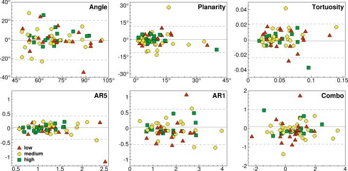
Materials and methods: Repeat scans of 61 participants, acquired as part of the Atherosclerosis Risk in Communities (ARIC) Carotid MRI substudy, were digitally segmented using automated 3D level set methods, relying on an operator only to select the branch endpoints and thresholds for the 3D lumen surface initialization. Geometric factors characterizing the 3D lumen geometry were then extracted automatically.
Results: Of 122 scans, 117 could be segmented within 5 minutes each, with 40% being of sufficiently high quality to require less than 2 minutes each. Irrespective of scan quality, geometric factors were found to be highly reproducible, with intraclass correlation coefficients (ICCs) typically above 0.9. The reconstructed lumen surfaces were reproducible to <0.3 mm on average, comparable to previous MRI-based reproducibility studies. Owing to the automated nature of the analysis, operator reliability was near-perfect (ICC >0.99), with lumen surface differences <0.1 mm.
Conclusion: The 3D geometry of the carotid bifurcation can be characterized rapidly and with a high degree of consistency, even for suboptimal image qualities. This bodes well for large-scale retrospective or prospective studies aimed at teasing out the influence of local vs. systemic risk factors for early atherosclerosis.
Copyright © 2011 Wiley-Liss, Inc.
Figures


References
-
- Slager CJ, Wentzel JJ, Gijsen FJ, et al. The role of shear stress in the generation of rupture-prone vulnerable plaques. Nature clinical practice. 2005;2(8):401–407. - PubMed
-
- Slager CJ, Wentzel JJ, Gijsen FJ, et al. The role of shear stress in the destabilization of vulnerable plaques and related therapeutic implications. Nature clinical practice. 2005;2(9):456–464. - PubMed
-
- Markl M, Wegent F, Zech T, et al. In-vivo Wall Shear Stress Distribution in the Carotid Artery: Effect of Bifurcation Geometry, Internal Carotid Artery Stenosis and Recanalization Therapy. Circ Cardiovasc Imaging - PubMed
Publication types
MeSH terms
Substances
Grants and funding
- N01 HC055016/HL/NHLBI NIH HHS/United States
- N01 HC055019/HL/NHLBI NIH HHS/United States
- N01-HC-55016/HC/NHLBI NIH HHS/United States
- U01 HL075572/HL/NHLBI NIH HHS/United States
- N01 HC055015/HL/NHLBI NIH HHS/United States
- N01-HC-55021/HC/NHLBI NIH HHS/United States
- N01 HC055020/HL/NHLBI NIH HHS/United States
- N01-HC-55019/HC/NHLBI NIH HHS/United States
- N01-HC-55015/HC/NHLBI NIH HHS/United States
- N01-HC-55020/HC/NHLBI NIH HHS/United States
- N01 HC055018/HL/NHLBI NIH HHS/United States
- N01-HC-55018/HC/NHLBI NIH HHS/United States
- N01-HC-55022/HC/NHLBI NIH HHS/United States
- N01 HC055021/HL/NHLBI NIH HHS/United States
- MOP-62934/CAPMC/ CIHR/Canada
- N01 HC055015/HC/NHLBI NIH HHS/United States
- N01 HC055022/HL/NHLBI NIH HHS/United States
- U01HL075572-01/HL/NHLBI NIH HHS/United States
LinkOut - more resources
Full Text Sources
Medical
