Review
. 2011 Sep 1;15(5):1179-93.
doi: 10.1089/ars.2011.3893. Epub 2011 May 9.

Claudins: control of barrier function and regulation in response to oxidant stress

Affiliations
Review

Claudins: control of barrier function and regulation in response to oxidant stress

Christian E Overgaard et al. Antioxid Redox Signal. .

Abstract

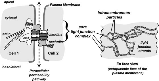
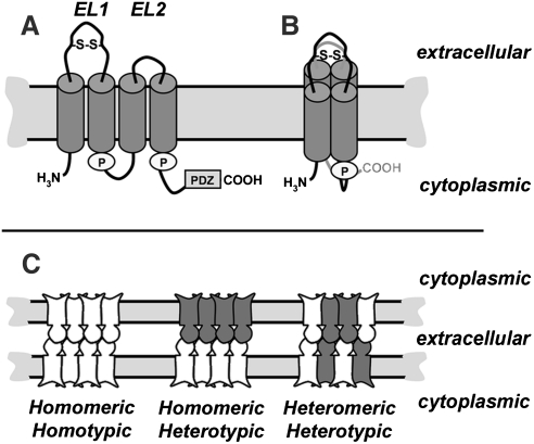
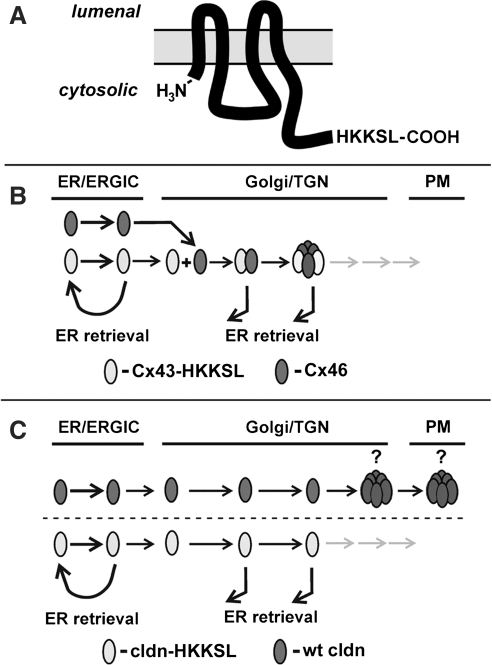
Claudins are a family of nearly two dozen transmembrane proteins that are a key part of the tight junction barrier that regulates solute movement across polarized epithelia. Claudin family members interact with each other, as well as with other transmembrane tight junction proteins (such as occludin) and cytosolic scaffolding proteins (such as zonula occludens-1 (ZO-1)). Although the interplay between all of these different classes of proteins is critical for tight junction formation and function, claudin family proteins are directly responsible for forming the equivalent of paracellular ion selective channels (or pores) with specific permeability and thus are essential for barrier function. In this review, we summarize current progress in identifying structural elements of claudins that regulate their transport, assembly, and function. The effects of oxidant stress on claudins are also examined, with particular emphasis on lung epithelial barrier function and oxidant stress induced by chronic alcohol abuse.


Figures

FIG. 1.
FIG. 1.
Core protein constituents of tight junctions. A diagram depicting the cell–cell contact site where tight junctions regulate paracellular permeability (dashed line and arrows). The core tight junction unit consists of transmembrane proteins (claudins, occludin) and scaffold proteins (ZO-1, ZO-2) which link claudins to the actin cytoskeleton. Head-to-head binding between claudins on adjacent cells provides the structural basis for the paracellular permeability barrier. Cartoon of an en face view which represents the appearance of tight junction strands as intramembraneous particles (circles) within the plasma membrane as visualized by freeze fracture electron microscopy. Each of the intramembraneous particles represents a complex containing several proteins, including claudins.
FIG. 2.
FIG. 2.
Homology between human claudins. A homology dendrogram was calculated by comparing the full length amino acid sequences of human claudins (hCldn) using ClustalW (19). Cldn-10a, cldn-10b, cldn-18, and cldn-18A2.1 are mRNA splice variants derived from the same gene product. The length of each branch line and point of divergence is proportional to the amount of inferred time of evolutionary divergence between different claudin genes (106). The dendrogram is restricted to human claudins known to be structural tight junction components; some claudin-related proteins in the Epithelial Membrane Protein family (68) were not included in this analysis.
FIG. 3.
FIG. 3.
Claudin secondary structure and classes of claudin-claudin interactions. (A) Line diagram showing key features of a typical claudin in the plane of the membrane, including the two extracellular loop (EL) domains, where EL1 contains a putative disulfide bond (S–S). Cylinders represent predicted transmembrane alpha helical domains. Also illustrated are two palmitoylation motifs (“P”) and the PDZ binding motif at the extreme C-terminus of the protein. (B) Proposed conformation of an individual claudin in the plane of the membrane, showing the four transmembrane alpha helical domains as a tightly packed complex. (C) Classes of claudin–claudin interactions within a tight junction strand. Depicted are tight junctions at sites where two cells are in contact composed of a single claudin (homomeric/homotypic) or multiple claudins (homomeric/heterotypic; heteromeric/heterotypic). Claudins can interact via head-to-head binding in the extracellular environment between adjacent cells (heterotypic interactions) and within the plane of the plasma membrane in the same cell (heteromeric interactions).
FIG. 4.
FIG. 4.
Use of a dilysine motif to produce ER-retained transmembrane proteins. (A) Placement of an HKKSL motif on the carboxyl-terminus of a tetraspan transmembrane protein, such as a claudin or connexin, acts as an ER-retention/retrieval signal (83). (B) For proteins that are heteromerically compatible to interact early in the secretory pathway, such as Cx43 and Cx46, the HKKSL-tagged protein acts as a dominant negative to prevent transport of wild-type proteins to the plasma membrane (PM). (C) By contrast, if an HKKSL-tagged claudin does not have the ability to inhibit the transport of a wild-type claudin, this indicates that the two claudins do not interact in the ER, Golgi intermediate compartment (ERGIC), or Golgi apparatus. (C) depicts the potential for claudin oligomerization occurring in either the trans Golgi network (TGN) or at the plasma membrane (PM). Immunofluorescence images corresponding to the models depicted in (B) and (C) are shown in Figure 5.
FIG. 5.
FIG. 5.
ER-retained claudins lack the ability to interact with untagged claudins. (A–L) Henrietta Lacks (HeLa) cells transfected to express either Cx43-HKKSL and Cx46 (A–C), cldn-4 and cldn-3 (D–F), cldn-4 and cldn-3-HKKSL (G–I), or cldn-4-HKKSL and cldn-3 (J–L) were fixed, permeabilized, and immunostained using mouse anti-Cx43 and rabbit anti-Cx46 (A–C) or mouse anti-cldn-4 and rabbit anti-cldn-3 (D–L) and Cy3-goat anti-mouse IgG and Cy2-goat anti-rabbit IgG. Arrowheads show areas where intracellular Cx43-HKKSL and Cx46 co-localize at the ER (A–C) and where cldn-3 and cldn-4 co-localize at the plasma membrane (D–F). There was little co-localization between the HKKSL tagged and untagged claudins (G–L). (M–O) MDCK cells were transiently transfected with myc-tagged cldn-4-HKKSL, fixed, permeabilized, and then immunostained using rabbit anti-cldn-3 and mouse anti-myc and the fluorescent secondary antibodies described above. Again, there was little co-localization between endogenously expressed cldn-3 and myc-cldn-4-HKKSL. Bar, 10 μm.
FIG. 6.
FIG. 6.
Changes in claudin expression in response to chronic alcohol ingestion. (A) Organization of alveolar epithelium, showing the organization of type I and type II alveolar epithelial cells in the terminal airspaces of the normal lung. The image presents an accurate representation of the relative size, distribution, and pattern of localization for alveolar epithelial cells in a healthy, adult lung. (B, C) Relative expression of cldn-1, −3, −4, −5, −7, and −18 by alveolar epithelial type II cells freshly isolated from rats (B) or cultured for 6 days to generate model type I cells (C). Rats were fed for 6 weeks using a calorically matched Liber-DeCarli liquid diet with 36% of the calories from maltodextrin (control) or ethanol (alcohol), harvested, and then analyzed by immunoblot, normalized to the level of GAPDH expression. Each claudin in the alcohol-fed experimental group was calculated as the mean+SE of six independent preparations, expressed as the percent change vs. control fed rats; p values are shown above pairs of bars. Chronic exposure to dietary alcohol had a significant effect on cldn-5 expression by type II cells; type I cells also showed decreases in several claudins, including cldn-1, cldn-7, and cldn-18. Data in (B, C) were modified from Fernandez, et al. (36).

References

    1. Amasheh S. Meiri N. Gitter AH. Schoneberg T. Mankertz J. Schulzke JD. Fromm M. Claudin-2 expression induces cation-selective channels in tight junctions of epithelial cells. J Cell Sci. 2002;115:4969–4976. - PubMed
    1. Amasheh S. Schmidt T. Mahn M. Florian P. Mankertz J. Tavalali S. Gitter AH. Schulzke JD. Fromm M. Contribution of claudin-5 to barrier properties in tight junctions of epithelial cells. Cell Tissue Res. 2005;321:89–96. - PubMed
    1. Anderson JM. Van Itallie CM. Physiology and function of the tight junction. Cold Spring Harb Perspect Biol. 2009;1:a002584. - PMC - PubMed
    1. Andras IE. Pu H. Deli MA. Nath A. Hennig B. Toborek M. HIV-1 Tat protein alters tight junction protein expression and distribution in cultured brain endothelial cells. J Neurosci Res. 2003;74:255–265. - PubMed
    1. Angelow S. Ahlstrom R. Yu AS. Biology of claudins. Am J Physiol Renal Physiol. 2008;295:F867–876. - PMC - PubMed

Publication types

LinkOut - more resources