. 2011 Apr;80(1):14-32.
doi: 10.1111/j.1365-2958.2011.07558.x. Epub 2011 Feb 10.

CaMtw1, a member of the evolutionarily conserved Mis12 kinetochore protein family, is required for efficient inner kinetochore assembly in the pathogenic yeast Candida albicans

Affiliations

CaMtw1, a member of the evolutionarily conserved Mis12 kinetochore protein family, is required for efficient inner kinetochore assembly in the pathogenic yeast Candida albicans

Babhrubahan Roy et al. Mol Microbiol. 2011 Apr.

Abstract

Proper assembly of the kinetochore, a multi-protein complex that mediates attachment of centromere DNA to spindle microtubules on each chromosome, is required for faithful chromosome segregation. Each previously characterized member of the Mis12/Mtw1 protein family is part of an essential subcomplex in the kinetochore. In this work, we identify and characterize CaMTW1, which encodes the homologue of the human Mis12 protein in the pathogenic budding yeast Candida albicans. Subcellular localization and chromatin immunoprecipitation assays confirmed CaMtw1 is a kinetochore protein. CaMtw1 is essential for viability. CaMtw1-depleted cells and cells in which CaMtw1 was inactivated with a temperature-sensitive mutation had reduced viability, accumulated at the G2/M stage of the cell cycle, and exhibited increased chromosome missegregation. CaMtw1 depletion also affected spindle length and alignment. Interestingly, in C. albicans, CaMtw1 and the centromeric histone, CaCse4, influence each other for kinetochore localization. In addition, CaMtw1 is required for efficient kinetochore recruitment of another inner kinetochore protein, the CENP-C homologue, CaMif2. Mis12/Mtw1 proteins have well-established roles in the recruitment and maintenance of outer kinetochore proteins. We propose that Mis12/Mtw1 proteins also have important co-dependent interactions with inner kinetochore proteins and that these interactions may increase the fidelity of kinetochore formation.


Figures

Figure 1
Figure 1
CaMtw1 is a kinetochore protein in C. albicans. (A) CaMtw1 co-localizes with the centromeric histone, CaCse4. Fixed cells of YJB10704 were stained with DAPI and immuno-stained with anti-CaCse4 antibody and anti-GFP (CaMtw1) antibodies (Bar, 2μm). (B and C) Live-cell time lapse imaging of CaMtw1-GFP in hyphal cells. Cells were imaged at 4 min intervals. Images shown are overlays of DIC and GFP images. The red and white arrowheads point to two CaMtw1-GFP foci, which after segregation were moving away from each other. One focus (red) was migrating back to the mother cell, and the other (white) was moving in the opposite direction (hyphal germ-tube). (D) Live-cell time-lapse imaging of CaMtw1-GFP in yeast-form cells. Cells were imaged at 4 min intervals. Images shown are overlays of DIC and GFP images. (E) Western blot analysis of whole cell extracts prepared from CAKS11 (MTW1/mtw1) and CAKS13 (MTW1-TAP/mtw1) cells with anti-protein A antibodies shows that CAKS13 expresses TAP-tagged CaMtw1 (predicted molecular weight of 57 kDa) protein whereas CAKS11 does not. (F) CaMtw1 localizes like a typical kinetochore protein. Fixed CAKS13 cells were stained by DAPI (DNA) and coimmunostained with anti-protein A (CaMtw1) and anti-tubulin (spindle microtubules) antibodies. Dot-like CaMtw1 signals were observed in the cells at different stages of cell cycle (Bar, 2μm). (G) PCR of ChIP assays reveal that the CaMtw1 is enriched within the CEN7 region of chromosome 7. Sheared chromatin fragments of CAKS13 strain (CaMtw1-TAP) were immunoprecipitated with anti-protein A antibodies. PCR reactions with primer pairs that amplify 178- to 292-bp regions spaced approximately every 1 kb between Orf19.6522 and Orf19.6524 of CEN7 region were used to amplify total DNA (SM) and immunoprecipitated DNA with anti-protein A antibodies and (+Ab) or beads only without (-Ab) antibody ChIP DNA fractions.
Figure 2
Figure 2
CaMtw1 is essential for C. albicans growth. Shutdown of CaMTW1 expression prevents CAKS12 (PCK1pr-MTW1/mtw1) strain growth on glucose media (YPD), whereas induction of CaMTW1 expression on succinate media allows CAKS12 cells to grow. Control strains BWP17 (wildtype) and CAKS11 (MTW1/mtw1) grow on both media. Plates were incubated at 30°C for 3 days.
Figure 3
Figure 3
CaMtw1 is needed for high fidelity chromosome segregation in C. albicans. (A) CAKS12 (PCK1pr-MTW1/mtw1) cell viability drops dramatically between 4 h and 6 h after repression of CaMTW1 expression on glucose. CAKS12 cells were grown overnight on succinate media, washed and shifted to glucose media. Cells were then harvested at 2 h intervals. Cell viability was calculated by plating diluted aliquots of culture at indicated time points on succinate plates and counting the number of colonies that appeared after incubating the plates for 3 days at 30°C. (B) Images of CAKS11 (MTW1/mtw1) and CAKS12 cells harvested at the indicated time points. (C) Distribution of unbudded (G1), small budded (S) and large budded (G2/M) cells of CAKS11 and CAKS12 after media shift from succinate to glucose. (D) FACS profiles of CAKS11 and CAKS12 cells incubated in glucose media for indicated time points and stained with propidium iodide. The X axis uses propidium iodide staining intensity as a measure of DNA content and the Y axis reports the relative number of cells.
Figure 4
Figure 4
Mtw1/Mis12 proteins have functional domains that are evolutionarily conserved in C. albicans. (A) Amino acid sequence alignment of two blocks of conserved amino acids present at the N terminal regions of the Mis12/Mtw1 proteins in different organisms: C. albicans CaMtw1, S. cerevisiae ScMtw1, S. pombe SpMis12, N. crassa NcMis12, C. neoformans CnMis12, D. hansenii DhMis12, and K. lactis KlMis12. The conserved glycine residue (asterisk) was mutated to glutamate, resulting in a temperature- sensitive mutation in strain CAKS14 (ts-mtw1/ mtw1). Multiple sequence alignment of the Mis12 sequences was performed by T-Coffee (http://www.ebi.ac.uk/Tools/t-coffee) and representation was done by ESPript 2.2 (http://espript.ibcp.fr/ESPript/cgi-bin/ESPript.cgi). (B) CAKS14 cells did not grow at 37°C, yet exhibited robust growth at 23°C and 30°C on YPDU plates for 2 days. Control parent strains BWP17 (MTW1/MTW1) and CAKS11 (MTW1/mtw1) cells grew normally at all three temperatures. (C) Flow cytometry profiles of CAKS11 and CAKS14 cells incubated at 37°C for indicated time points and stained with propidium iodide. The X axis uses propidium iodide staining intensity as a measure of DNA content and the Y axis reports the number of cells.
Figure 5
Figure 5
CaMtw1 depletion causes defects in spindle alignment, position, and length. (A) BWP17 (MTW1/MTW1) and CAKS12 (PCK1pr-MTW1/mtw1) cells (grown in glucose media for 7 h at 30°C) were fixed and stained with anti-tubulin (spindle) antibodies and DAPI (DNA). (Bar, 2μm). The percent of cells showing different spindle morphologies are mentioned below every image. (B) Percentage of properly and improperly aligned spindles were counted in BWP17 (71 cells) and CAKS12 (117 cells). (C) Mitotic spindle lengths were measured by Zeiss LSM software in BWP17 (70 cells) and CAKS12 (131 cells). Percentage of cells carrying spindles of indicated length is represented.
Figure 6
Figure 6
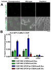
CaCse4 influences localization of CaMtw1 at the centromere. (A) GFP and merged GFP-DIC microscopy images showing CaMtw1-GFP signals in YJB11482 (CSE4/cse4) (wild-type) and YJB11483 (PCK1pr-CSE4/cse4) under conditions that repress (glucose) or induce (succinate) expression of the PCK1 promoter. (Bar, 5μm). (B) ChIP assays were performed on strains YJB11482 (CSE4/cse4) and YJB11483 (PCK1pr-CSE4/cse4) grown in glucose (repressing condition for PCK1pr) or succinate (inducing condition for PCK1pr) for 6h, using anti-GFP antibodies and primer pairs that amplify the central regions of CEN5 (JB3924/JB3925) and CEN7 (JB3993/JB3994). Amplification from the LEU2 ORF (JB4165/ JB4166) was also performed to detect background DNA elution in the ChIP assays. qPCR of total DNA and with (+) or without (-) antibody ChIP DNA fractions were performed. Enrichment of CaMtw1-GFP was calculated as a percentage of the total chromatin input. Shown are the averages of two representative biological replicates ± SEM. t-tests were used to compare CaMtw1-GFP recruitment at the central CEN5 and CEN7 regions in YJB11482 and YJB11483 in glucose (p<0.01) and succinate (p<0.01) (Indicated by *).
Figure 7
Figure 7
CaCse4 localization at the centromere is affected by CaMtw1: (A) GFP and merged GFP-DIC microscopy images showing CaCse4-GFP signals in YJB11553 (MTW1/mtw1) and YJB11554 (PCK1pr-MTW1/mtw1) in repressing (glucose) and inducing (succinate) conditions for the PCK1 promoter. (Bar, 5μm) (B) ChIP assays were performed on strains CAKS12 (grown in glucose for 6h, where PCK1pr-CaMTW1 is repressed) and CAKS11 (with wild-type CaMTW1 expressed) using anti-CaCse4 antibodies and primer pairs that amplify the central regions of CEN5 (CACH5F1/CACH5R1) and CEN7 (nCEN7-3/nCEN7-4). qPCR of total DNA and with (+) or without (-) antibody ChIP DNA fractions were performed. Enrichment of CaCse4 at the centromere was calculated as a percentage of the total chromatin input. Amplification from LEU2 ORF (nLeu2- 1/nLeu2-2) was also performed to detect the background DNA elution in the ChIP assays. t-tests were used to compare CaCse4 recruitment at the central CEN5 and CEN7 region in YJB11553 and YJB11554 in glucose (p<0.01) (indicated by *). (C) CAK11 (MTW1/mtw1) and CAKS14 (ts-mtw1/mtw1) cells (grown at 23°C and 37°C) were fixed and stained with anti-CaCse4 antibodies and DAPI (nucleus). (Bar, 5μm). (D) Standard ChIP assays were performed on strains CAKS14 and CAKS11 (grown at 37°C) using anti-CaCse4 antibodies and primers that amplify the central regions of CEN5 (CACH5F1/CACH5R1) and CEN7 (nCEN7-3/nCEN7-4). PCR of total DNA and with (+) or without (-) antibody ChIP DNA fractions were performed. QPCR amplification from LEU2 ORF (nLeu2-1/nLeu2-2) was also done to detect the background DNA elution in the ChIP assays. Enrichment of CaCse4 at the centromere was calculated as a percentage of the total chromatin input. t-tests were used to compare CaCse4 recruitment at the central CEN5 and CEN7 region in CAKS11 and CAKS14 at 37° C (p<0.005) (indicated by *).
Figure 8
Figure 8
CaMif2 localization at the centromere is affected by CaMtw1: (A) GFP and merged GFP-DIC microscopy images showing CaMif2-GFP signals in YJB12118 (MTW1/mtw1) and YJB12119 (PCK1pr-MTW1/mtw1) in repressing (glucose) and inducing (succinate) conditions for the PCK1 promoter (Bar, 5μm). (B) ChIP assays were performed on strains YJB12119 [grown in succinate (CaMtw1 is overexpressed) and glucose for 6 h (CaMtw1 is repressed)] and YJB12118 (with wild-type CaMTW1 expressed) using anti-GFP antibodies and primer pairs that amplify the central regions of CEN5 (JB3924/JB3925) and CEN7 (JB3993/JB3994). qPCR of total DNA and with (+) or without (-) antibody ChIP DNA fractions were performed. Enrichment of CaMif2 at the centromere was calculated as a percentage of the total chromatin input. Amplification from LEU2 ORF (JB4165/ JB4166) was also performed to detect the background DNA elution in the ChIP assays. t-tests were used to compare CaMif2 recruitment at the central CEN5 and CEN7 region in YJB12118 and YJB12119 in glucose (p<0.001) and in succinate (p<0.05) (indicated by *). (C) ChIP assays were performed on strains YJB12118 and YJB12176 (grown both at 23°C and 37°C for 2 h) using anti-GFP antibodies and primers that amplify the central regions of CEN5 (JB3924/JB3925) and CEN7 (JB3993/JB3994). PCR of total DNA and with (+) or without (-) antibody ChIP DNA fractions were performed. QPCR amplification from LEU2 ORF (JB4165/ JB4166) was also done to detect the background DNA elution in the ChIP assays. Enrichment of CaMif2 at the centromere was calculated as a percentage of the total chromatin input. t-tests were used to compare CaMif2 recruitment at the central CEN5 and CEN7 region in YJB12118 and YJB12176 at 37° C (p< 0.001) (indicated by *).

Similar articles

Cited by

References

    1. Allshire RC, Karpen GH. Epigenetic regulation of centromeric chromatin: old dogs, new tricks. Nat Rev Genet. 2008;9:923–37. - PMC - PubMed
    1. Bachewich C, Nantel A, Whiteway M. Cell cycle arrest during S or M phase generates polarized growth via distinct signals in Candida albicans. Mol Microbiol. 2005;57:942–959. - PubMed
    1. Baum M, Clarke L. Fission yeast homologs of human CENP-B have redundant functions affecting cell growth and chromosome segregation. Mol Cell Biol. 2000;20:2852–2864. - PMC - PubMed
    1. Baum M, Sanyal K, Mishra PK, Thaler N, Carbon J. Formation of functional centromeric chromatin is specified epigenetically in Candida albicans. Proc Natl Acad Sci U S A. 2006;103:14877–14882. - PMC - PubMed
    1. Berman J. Morphogenesis and cell cycle progression in Candida albicans. Curr Opin Microbiol. 2006;9:595–601. - PMC - PubMed

Publication types

MeSH terms

LinkOut - more resources