. 2011 Jan;11(56):7-17.

The vitamin D receptor: a tumor suppressor in skin

Affiliations

The vitamin D receptor: a tumor suppressor in skin

Daniel David Bikle. Discov Med. 2011 Jan.

Abstract

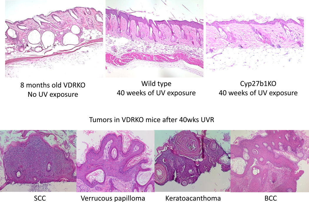
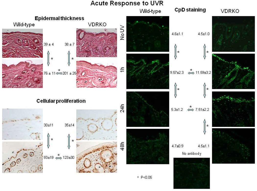
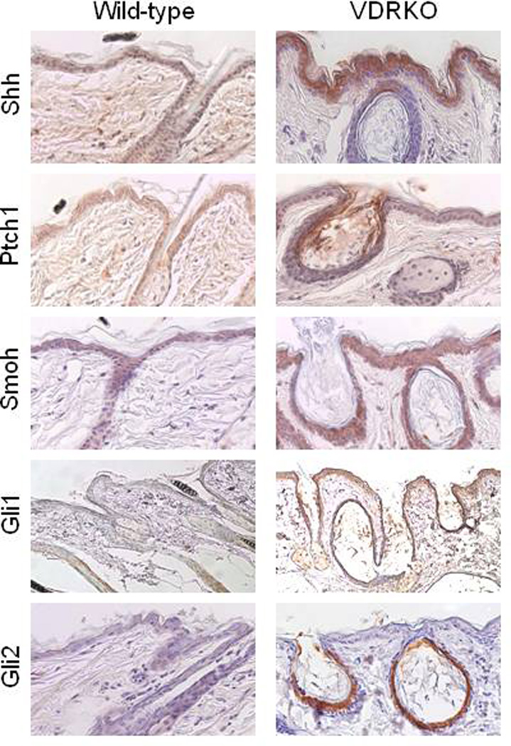
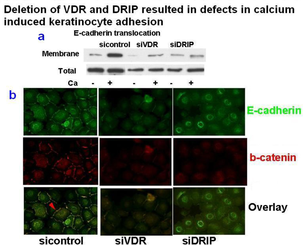
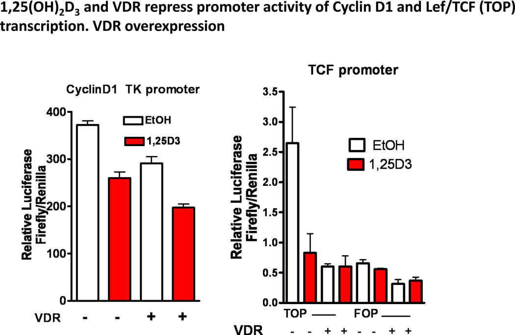
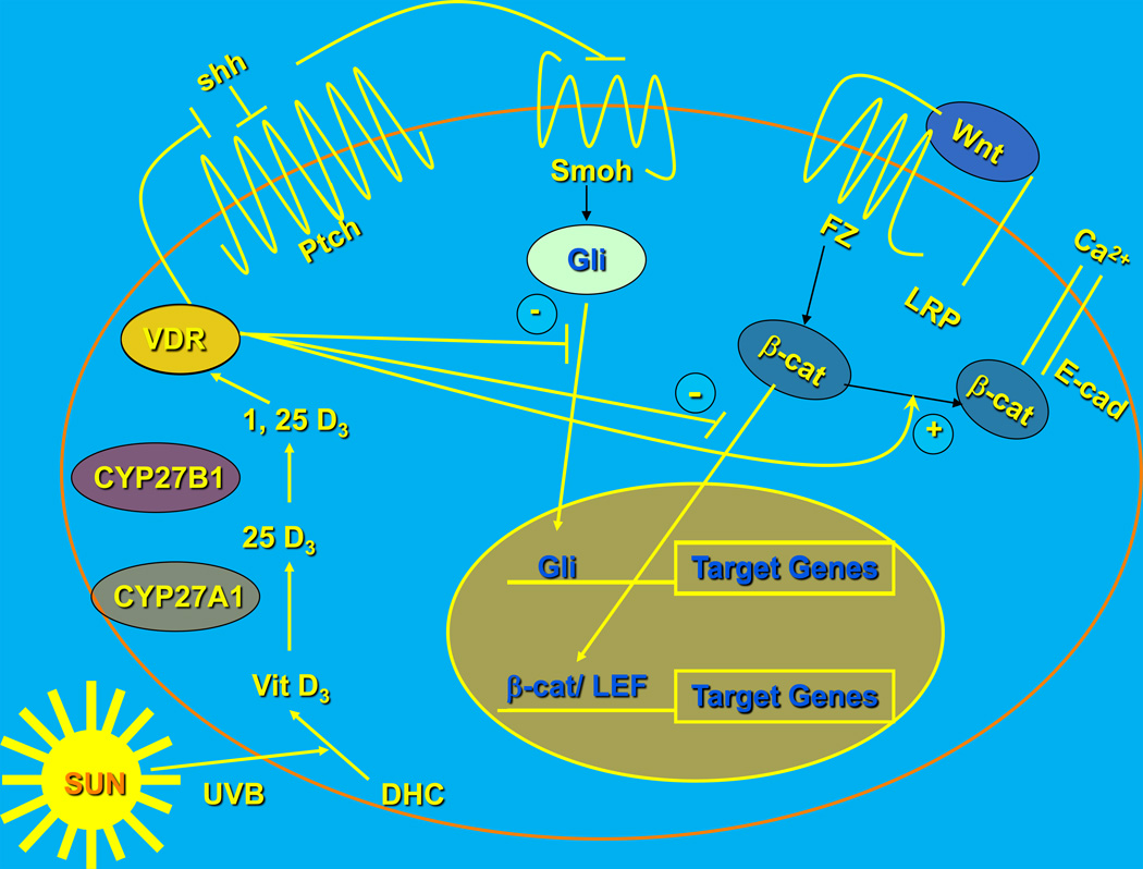
Epidemiologic evidence supporting a major chemopreventive role for vitamin D in various malignancies is strong. Likewise the use of the active metabolite of vitamin D, 1,25(OH)(2)D(3), and its analogs to prevent and/or treat a wide variety of malignancies in animals is well established. The evidence has been less compelling for epidermal carcinogenesis perhaps because the same agent that produces vitamin D in the skin, UVB radiation (UVR), is also the same agent that results in most epidermal malignancies. However, recent studies indicate that the role of vitamin D and its receptor (VDR) in protecting against the development of epidermal tumors deserves a closer look. One such study found mice lacking the VDR were quite sensitive to epidermal tumor formation following the administration of the carcinogen DMBA. A more recent study showed that these mice were similarly more sensitive to tumor formation following UVR, results we have confirmed. The epidermis of the VDR null mouse is hyperproliferative with gross distortion of hair follicles, structures that may provide the origin for the tumors found in the skin following such treatment. Two interacting pathways critical for epidermal and hair follicle function, beta-catenin and hedgehog (Hh), result in epidermal tumors when they are activated abnormally. Thus, we considered the possibility that loss of VDR predisposes to epidermal tumor formation by activation of either or both beta-catenin and Hh signaling. We determined that all elements of the Hh signaling pathway are upregulated in the epidermis and utricles of the VDR null mouse, and that 1,25(OH)(2)D(3) suppresses the expression of these elements in normal mouse skin. In addition we observed that the transcriptional activity of beta-catenin was increased in keratinocytes lacking the VDR. These results lead us to the hypothesis that the VDR with its ligand 1,25(OH)(2)D(3) functions as a tumor suppressor with respect to epidermal tumor formation in response to UVR by regulating Hh and beta-catenin signaling.


Conflict of interest statement

Disclosure

The authors report no conflicts of interest.

Figures

Figure 1
Figure 1. The Sonic hedgehog (Shh) signaling pathway
In the absence of Shh, Ptch 1 suppresses signaling by smoothened (Smoh). Binding of Shh to Ptch 1 relieves this inhibition. Activation of Smoh leads to the activation and translocation of transcription factors of the Gli family into the nucleus, with subsequent changes in gene expression.
Figure 2
Figure 2. The canonical wnt signaling pathway
Wnts bind to their frizzled receptors (FZ) and coreceptors LRP in the membrane. This binding can be blocked by dickkopf (Dkk) or soluble frizzled related proteins (sFRP). Activation of FZ by wnt results in phosphorylation of disheveled (Dvl) which induces the disruption of the axin/APC/GSK-3b complex and recruitment of axin to the membrane. This complex, when active, phosphorylates β-catenin, leading to its proteosomal degradation. However, following wnt stimulation, β-catenin is no longer degraded and can enter the nucleus where in combination with members of the LEF/TCF family can induce expression of its target genes such as cyclin D1. β-catenin also binds to the E-cadherin complex in the plasma membrane, a complex stabilized by calcium, where it may play a role in differentiation.
Figure 3
Figure 3. Tumors in the VDRKO mice following UVR
In the upper panels VDRKO mice not exposed to UVR but of comparable age to the wildtype and CYP27B1KO mice exposed to 40 weeks of UVR are shown. No tumors were found in these mice. In contrast all VDRKO mice developed tumors during UVR. Representative tumor types found in these mice are shown in the lower panels.
Figure 4
Figure 4. Acute response of VDRKO and wildtype mouse skin to UVR: proliferation and CPD formation
The mice were irradiated with 1 dose of 400mJ/cm2 UVB, and their skin obtained prior to and up to 48 hours after UVR for measurement of epidermal thickness, immunostaining for PCNA, a marker of proliferation, and CPDs, a measure of DNA damage. Although the rates of proliferation and CPD formation increased in both wildtype and VDR null mice after UVR, by 48 hours the epidermis was thicker with significantly greater numbers of proliferating keratinocytes and more CPDs in the VDR null mouse epidermis. The numbers represent mean +/− SD of 6 images per mouse and 3 mice/group.
Figure 5
Figure 5. The skin of adult VDR null mice express an increase in components of the Hh pathway
Immunohistochemical localization of Shh, Ptch1, Smoh, Gli1, and Gli2 was performed in 11-week wildtype and VDR null mice. Unlike the wildtype littermates, VDR null mouse skin continues to overexpress elements of the Hh pathway.
Figure 6
Figure 6. 1,25(OH)2D3 suppresses Shh expression
Mice with floxed VDR were bred with mice transgenic for Cre recombinase driven by the K14 promoter to provide epidermal specific deletion of VDR. Epidermal sheets from neonatal mice in which VDR was deleted (Cre) and their control littermates (no Cre) were cultured for 24 hours in media containing 10nM 1,25(OH)2D3 or vehicle (ethanol) ) for 24 hours. L19 was used to normalize the polymerase chain reaction (PCR) data. The data are expressed as mean +/− SD of triplicates. 1,25(OH)2D3 significantly inhibited expression of Shh. * P<0.01.
Figure 7
Figure 7. VDR and its coactivator DRIP205 regulate membrane formation of the E-cadherin/β-catenin complex
Human foreskin keratinocytes were transfected with siRNA for VDR, DRIP205, and non-target siRNA as control in low calcium media (0.03mM calcium) for 3 days. The cells were then treated with high calcium (1.2mM) for 5 minutes to induce cell adhesion. The translocation of E-cadherin and β-catenin to the membrane was observed by Western blot analysis and immunohistochemistry. When keratinocytes were treated with high calcium, cell adhesion was induced. Both E-cadherin and β-catenin were translocated to the membrane to form adherens junctions (red arrow). When VDR and DRIP expressions were silenced using siRNA (siVDR and siDRIP, respectively), this adhesion was abolished as shown by lack of translocation of siVDR, siDRIP for both E-cadherin and β-catenin. E-cadherin expression was reduced by silencing of VDR and DRIP expression. The data shown are representative of three experiments with comparable results.
Figure 8
Figure 8. Inhibition of the cyclin D1 promoter and an LEF/TCF promoter construct by VDR and 1,25(OH)2D3
Keratinocytes were transfected with luciferase constructs linked to a cyclin D1 TK promoter, an LEF/TCF promoter (TOP Glow), or its mutated control (FOP Glow). The cells were cotransfected with CMV hVDR vector (+) to overexpress VDR, or control pcDNA vector (−), and control renilla luciferase (pRL-TK). The cells were treated with 1,25(OH)2D3 or vehicle. The firefly and renilla luciferase activities were measured by dual luciferase kit, and the ratios of these luciferases were calculated. Both VDR and 1,25(OH)2D3 inhibited the cyclin D1 and LEF/TCF promoter constructs with little effect on the mutated LEF/TCF promoter construct. The error bars enclose mean +/− SD of triplicate cultures.
Figure 9
Figure 9. Working model for the regulation of Hh and β-catenin signaling in keratinocytes by 1,25(OH)2D3 and VDR: their role as tumor suppressors
The keratinocyte is capable of making its own 1,25(OH)2D3 from the vitamin D3 produced from 7-dehydrocholesterol (DHC) under the influence of UVB, as it has both CYP27A1 (which converts vitamin D3 to 25OHD3) and CYP27B1 (which converts 25OHD3 to 1,25(OH)2D3). The VDR with or without its ligand suppresses Shh expression. It binds β-catenin and induces E-cadherin expression reducing the amount of β-catenin available for binding to LEF1. It may also have a direct inhibitory action on Gli transcriptional activity by direct binding similar to its binding to β-catenin (postulated, not demonstrated). These actions regulate transcriptional activity of both Gli and β-catenin, reducing their proliferative actions and limiting their ability to induce tumors in the skin.

References

    1. Aszterbaum M, Epstein J, Oro A, Douglas V, Leboit PE, Scott MP, Epstein EH., Jr Ultraviolet and ionizing radiation enhance the growth of BCCs and trichoblastomas in patched heterozygous knockout mice. Nat Med. 1999;5(11):1285–1291. - PubMed
    1. Aszterbaum M, Rothman A, Johnson RL, Fisher M, Xie J, Bonifas JM, Zhang X, Scott MP, Epstein EH., Jr Identification of mutations in the human PATCHED gene in sporadic basal cell carcinomas and in patients with the basal cell nevus syndrome. J Invest Dermatol. 1998;110(6):885–888. - PubMed
    1. Barnfield PC, Zhang X, Thanabalasingham V, Yoshida M, Hui CC. Negative regulation of Gli1 and Gli2 activator function by Suppressor of fused through multiple mechanisms. Differentiation. 2005;73(8):397–405. - PubMed
    1. Bienz M. beta-Catenin: a pivot between cell adhesion and Wnt signalling. Curr Biol. 2005;15(2):R64–R67. - PubMed
    1. Bijlsma MF, Spek CA, Zivkovic D, Van De Water S, Rezaee F, Peppelenbosch MP. Repression of smoothened by patched-dependent (pro-)vitamin D3 secretion. PLoS Biol. 2006;4(8):e232. - PMC - PubMed