

Dihydroisoxazole analogs for labeling and visualization of catalytically active transglutaminase 2
- PMID: 21276939
- PMCID: PMC3073585
- DOI: 10.1016/j.chembiol.2010.11.004
Dihydroisoxazole analogs for labeling and visualization of catalytically active transglutaminase 2
Abstract
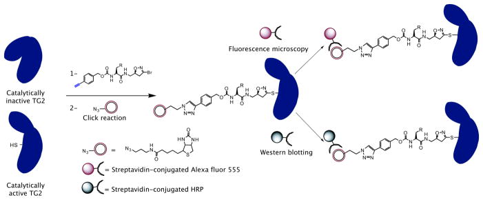
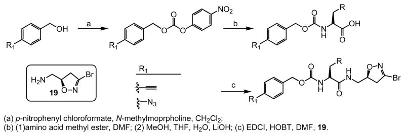
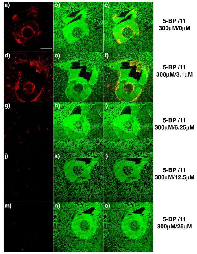
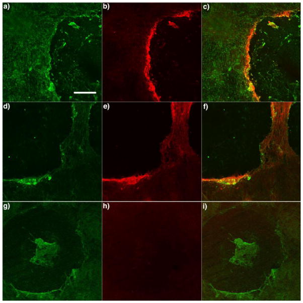
We report the synthesis and preliminary characterization of "clickable" inhibitors of human transglutaminase 2 (TG2). These inhibitors possess the 3-halo-4,5-dihydroisoxazole warhead along with bioorthogonal groups such as azide or alkyne moieties that enable subsequent covalent modification with fluorophores. Their mechanism for inhibition of TG2 is based on halide displacement, resulting in the formation of a stable imino thioether. Inhibition assays against recombinant human TG2 revealed that some of the clickable inhibitors prepared in this study have comparable specificity as benchmark dihydroisoxazole inhibitors reported earlier. At low micromolar concentrations they completely inhibited transiently activated TG2 in a WI-38 fibroblast scratch assay and could subsequently be used to visualize the active enzyme in situ. The potential use of these inhibitors to probe the role of TG2 in celiac sprue as well as other diseases is discussed.
Copyright © 2011 Elsevier Ltd. All rights reserved.
Figures

Similar articles
-
Substrates, inhibitors, and probes of mammalian transglutaminase 2.Anal Biochem. 2020 Feb 15;591:113560. doi: 10.1016/j.ab.2019.113560. Epub 2019 Dec 24. Anal Biochem. 2020. PMID: 31874171 Free PMC article. Review.
-
Structure-activity relationship analysis of the selective inhibition of transglutaminase 2 by dihydroisoxazoles.J Med Chem. 2006 Dec 14;49(25):7493-501. doi: 10.1021/jm060839a. J Med Chem. 2006. PMID: 17149878 Free PMC article.
-
Discovery of potent and specific dihydroisoxazole inhibitors of human transglutaminase 2.J Med Chem. 2014 Nov 13;57(21):9042-64. doi: 10.1021/jm501145a. Epub 2014 Oct 31. J Med Chem. 2014. PMID: 25333388 Free PMC article.
-
Chemistry and biology of dihydroisoxazole derivatives: selective inhibitors of human transglutaminase 2.Chem Biol. 2005 Apr;12(4):469-75. doi: 10.1016/j.chembiol.2005.02.007. Chem Biol. 2005. PMID: 15850984
-
Irreversible inhibitors of tissue transglutaminase.Adv Enzymol Relat Areas Mol Biol. 2011;78:415-47. doi: 10.1002/9781118105771.ch10. Adv Enzymol Relat Areas Mol Biol. 2011. PMID: 22220480 Review. No abstract available.
Cited by
-
Application of a Fluorescence Anisotropy-Based Assay to Quantify Transglutaminase 2 Activity in Cell Lysates.Int J Mol Sci. 2022 Apr 19;23(9):4475. doi: 10.3390/ijms23094475. Int J Mol Sci. 2022. PMID: 35562866 Free PMC article.
-
Bidirectional histone monoaminylation dynamics regulate neural rhythmicity.Nature. 2025 Jan;637(8047):974-982. doi: 10.1038/s41586-024-08371-3. Epub 2025 Jan 8. Nature. 2025. PMID: 39779849 Free PMC article.
-
An unprecedented dual antagonist and agonist of human Transglutaminase 2.Bioorg Med Chem Lett. 2015 Nov 1;25(21):4922-4926. doi: 10.1016/j.bmcl.2015.05.006. Epub 2015 May 15. Bioorg Med Chem Lett. 2015. PMID: 26004580 Free PMC article.
-
Substrates, inhibitors, and probes of mammalian transglutaminase 2.Anal Biochem. 2020 Feb 15;591:113560. doi: 10.1016/j.ab.2019.113560. Epub 2019 Dec 24. Anal Biochem. 2020. PMID: 31874171 Free PMC article. Review.
-
Elevated transglutaminase 2 activity is associated with hypoxia-induced experimental pulmonary hypertension in mice.ACS Chem Biol. 2014 Jan 17;9(1):266-75. doi: 10.1021/cb4006408. Epub 2013 Nov 5. ACS Chem Biol. 2014. PMID: 24152195 Free PMC article.
References
-
- Akimov SS, Belkin AM. Cell surface tissue transglutaminase is involved in adhesion and migration of monocytic cells on fibronectin. Blood. 2001;98:1567–1576. - PubMed
-
- Balklava Z, Verderio E, Collighan R, Gross S, Adams J, Griffin M. Analysis of tissue transglutaminase function in the migration of swiss 3T3 fibroblasts - The active-state conformation of the enzyme does not affect cell motility but is important for its secretion. J Biol Chem. 2002;277:16567–16575. - PubMed
-
- Case A, Ni J, Yeh LA, Stein RL. Development of a mechanism-based assay for tissue transglutaminase - results of a high-throughput screen and discovery of inhibitors. Anal Biochem. 2005;338:237–244. - PubMed
Publication types
MeSH terms
Substances
Grants and funding
LinkOut - more resources
Full Text Sources
Other Literature Sources
