. 2011 Feb 11;88(2):138-49.
doi: 10.1016/j.ajhg.2010.12.012. Epub 2011 Feb 3.

Mutations in prickle orthologs cause seizures in flies, mice, and humans

Affiliations

Mutations in prickle orthologs cause seizures in flies, mice, and humans

Hirotaka Tao et al. Am J Hum Genet. .

Abstract

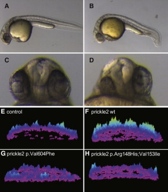
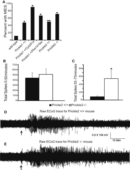
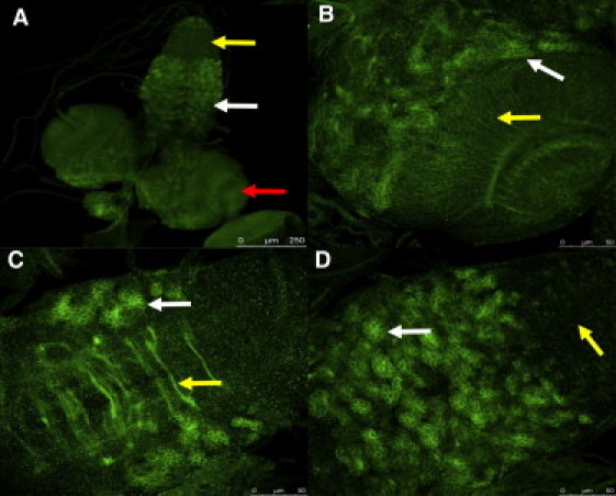
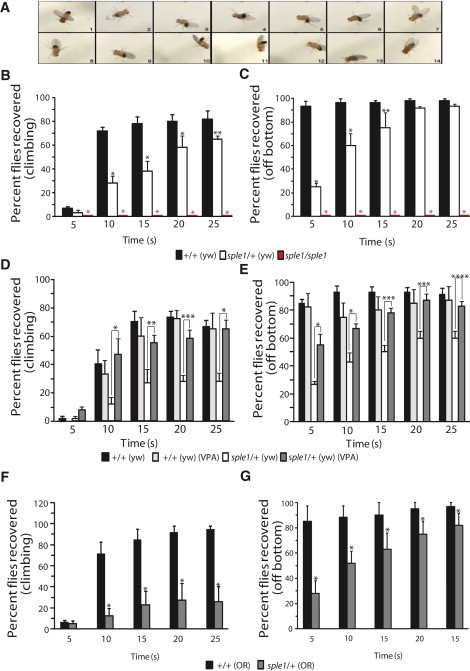
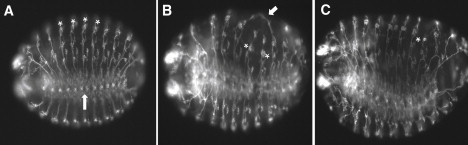
Epilepsy is heritable, yet few causative gene mutations have been identified, and thus far no human epilepsy gene mutations have been found to produce seizures in invertebrates. Here we show that mutations in prickle genes are associated with seizures in humans, mice, and flies. We identified human epilepsy patients with heterozygous mutations in either PRICKLE1 or PRICKLE2. In overexpression assays in zebrafish, prickle mutations resulted in aberrant prickle function. A seizure phenotype was present in the Prickle1-null mutant mouse, two Prickle1 point mutant (missense and nonsense) mice, and a Prickle2-null mutant mouse. Drosophila with prickle mutations displayed seizures that were responsive to anti-epileptic medication, and homozygous mutant embryos showed neuronal defects. These results suggest that prickle mutations have caused seizures throughout evolution.


Figures

Figure 1
Figure 1
Zebrafish Prickle Mutations Orthologous to Those Identified in Human Epilepsy Patients Show Altered Activity in CE and Calcium Release (A–D) Morphological phenotypes at 28 hpf. Compared to wild-type uninjected embryos (A), prickle2-injected embryos (B) display a shorter A-P axis and a kinky tail. The lateral view is shown, and anterior is to the left. Moreover, prickle2-injected embryos (D) show defects in eye and forebrain patterning at 28 hpf. (C) Uninjected embryo. Ventral view. The numbers of embryos with defects in each RNA-injected group are shown in Table 2. (E–H) Surface plots of calcium release activity were generated from images of live zebrafish embryos. The height and color of the peaks indicates the number of calcium fluxes observed over the course of the experiment; the embryos are oriented in a lateral position. (E) A control injected embryo. (F) An embryo overexpressing wild-type prickle2 RNA. (G) An embryo overexpressing prickle2Val605Phe-mutant-encoding RNA. (H) An embryo overexpressing prickle2 Arg148His; Val153Ile-mutant-encoding RNA.
Figure 2
Figure 2
Mouse Models Deficient for the Prickle Genes Show an Increased Seizure Phenotype (A) Maximal electroshock seizure threshold (MEST) in wild-type mice versus mice deficient for the prickle genes. Prickle1+/−, Prickle1+/Cys251X, Prickle1+/Phe141Ser, Prickle2+/−, and Prickle2−/− mice show a significantly lower MEST than wild-type mice at 7 mA (p < 0.001, ∗∗p < 0.005; wild-type n = 43, Prickle1+/− n = 23, Prickle1+/Cys251X n = 10, Prickle1+/Phe141Ser n = 12, Prickle2+/− n = 14, and Prickle2−/− n = 7). (B and C) Total number of electrographic spikes recorded from electrocorticography (ECoG) in response to pentylenetetrazol (PTZ) injection for the first 50 min after injection (B) and the last 25 min of recording after injection (C). During the first 50 min after injection, there is no difference in the total number of spikes between wild-type and Prickle2−/− mice. However, there is a significant difference seen during the last 25 min of recording: Prickle2−/− mice had significantly more seizures than their wild-type counterparts. p = 0.039 as determined by the Student's t test; wild-type n = 7, Prickle2−/− n = 6. Error bars represent SEM. (D and E) Representative ECoG traces for wild-type and Prickle2−/− mice upon stimulation with PTZ. PTZ was injected (black arrow) after 15 min of baseline recording. ECoG was recorded for 75 min after injection. A wild-type raw trace recording is shown in (D). A Prickle2−/− raw trace recording is shown in (E). In comparison to wild-type mice, Prickle2−/− mice continue to seize for prolonged periods after injection.
Figure 3
Figure 3
Prickle Is Expressed Widely in the Drosophila CNS A Prickle-specific polyclonal antibody was used for visualization of Prickle in the crawling third-instar larval CNS. Most neurons of the larval CNS persist through metamorphosis and join groups of newly born adult-specific neurons to form the adult CNS. (A) Prickle is localized in the optic lobes (one lobe is indicated by the red arrow), as well as central brain structures between the lobes, and to part of the ventral segmental ganglia (white arrow). High-level staining is not seen in ventral segmental ganglia that correspond to the abdominal segments (yellow arrow). (B) A higher magnification of the optic lobe region of the brain shows Prickle staining in clusters of bundled neurons (white arrow) as well as neuronal projections spanning the optic lobe (yellow arrow). (C) A higher magnification of the ventral segmental ganglia shows Prickle staining in the commisures that cross the ganglia (yellow arrow) as well as in clusters of neurons similar to those seen in the optic lobe regions of the brain (white arrow). (D) A higher magnification of the surface of the ventral segmental ganglia shows Prickle staining in clusters of neurons (white arrow); note that the ventral segmental ganglia corresponding to the abdominal segments is largely devoid of high Prickle staining (yellow arrow).
Figure 4
Figure 4
Drosophila that are Homozygous or Heterozygous for the prickle mutation pksple1Are Predisposed to Seizures, and Seizures are Diminished by Treatment with Valproic Acid (A) Seizure in a pksple1 fly. Photos of typical homozygous pksple1-seizing fly were captured every 0.5 s after the bang assay. All control flies have already climbed up the side of the vial for the time period presented. For a live side-by-side comparison of control and pksple1 flies, see Movies S5–S9. (B) pksple1 homozygous flies and pksple1 heterozygous flies in the yw67 (yw) background [pksple1/+ (yw)] have significantly impaired seizure recovery when they are compared to same-aged control yw67 [+/+(yw)] flies. p < 0.001, ∗∗p = 0.039. (C) pksple1 homozygous and heterozygous flies have significantly impaired recovery off the bottom in comparison to same-aged control +/+(yw) flies. p < 0.001, ∗∗p = 0.001. (D) The addition of valproic acid to fly food significantly diminishes seizures in pksple1 heterozygotes [pksple1/+ (yw)] and returns climbing ability to that of control +/+(yw) flies. p < 0.001, ∗∗p = 0.002, ∗∗∗p = 0.001. (E) The addition of valproic acid to fly food significantly recovers the ability of the flies to right themselves off the bottom of the vial and climb. p = 0.002, ∗∗p = 0.010, ∗∗∗p = 0.001, ∗∗∗p = 0.005. (F and G) pksple1 heterozygous flies are bang sensitive in multiple backgrounds. (F) pksple1 heterozygous flies in the Oregon-R (OR) background [sple1/+(OR)] have significantly impaired seizure recovery in comparison to same-aged wild-type Oregon-R [+/+ (OR)] flies. p = 0.007. (G) sple1/+(OR) heterozygous flies have significantly impaired recovery off bottom in comparison to same-aged wild-type +/+(OR) flies. p = 0.007. For all experiments, wild-type n = 60 and pksple1 n = 60. Error bars represent standard error of the mean; the data were statistically analyzed with the Chi-square test.
Figure 5
Figure 5
Neuronal Defects Are Observed in prickle Mutant Embryos Images (20×) of control 14–16 hr yw67 embryos (A) are compared to two 14–16 hr pksple1homozygous mutant embryos (B and C) that have been stained with the 22C10 antibody so that PNS neurons are visualized. Note the location of chordotonal organs (asterisks in A) as well as the location of the ventral nerve chord (upward arrow in A) in the control embryo. For the mutant embryo in (B), note the aberrant neuronal processes that have joined (arrow) as well as the abnormal location of two chordotonal organs (asterisks). For the mutant embryo in (C), note the overall disorganized PNS staining pattern, as well as the improperly positioned neurons associated with the chordotonal organ in abdominal segment 6 (asterisk). Anterior is to the left, and ventral is down but slightly rotated toward the viewer.

Comment in

References

    1. Frankel W.N. Genetics of complex neurological disease: challenges and opportunities for modeling epilepsy in mice and rats. Trends Genet. 2009;25:361–367. - PMC - PubMed
    1. Bassuk A.G., Wallace R.H., Buhr A., Buller A.R., Afawi Z., Shimojo M., Miyata S., Chen S., Gonzalez-Alegre P., Griesbach H.L. A homozygous mutation in human PRICKLE1 causes an autosomal-recessive progressive myoclonus epilepsy-ataxia syndrome. Am. J. Hum. Genet. 2008;83:572–581. - PMC - PubMed
    1. Goldschmidt R.B. A study of a spontaneous mutation. University of California Publications in Zoology. 1945;49:503–504.
    1. Gubb D., Green C., Huen D., Coulson D., Johnson G., Tree D., Collier S., Roote J. The balance between isoforms of the prickle LIM domain protein is critical for planar polarity in Drosophila imaginal discs. Genes Dev. 1999;13:2315–2327. - PMC - PubMed
    1. Veeman M.T., Slusarski D.C., Kaykas A., Louie S.H., Moon R.T. Zebrafish prickle, a modulator of noncanonical Wnt/Fz signaling, regulates gastrulation movements. Curr. Biol. 2003;13:680–685. - PubMed

Publication types

MeSH terms