. 2011 Apr;21(4):195-201.
doi: 10.1016/j.tcb.2010.12.006. Epub 2011 Jan 27.

The Three Musketeers of Autophagy: phosphorylation, ubiquitylation and acetylation

Affiliations

The Three Musketeers of Autophagy: phosphorylation, ubiquitylation and acetylation

David G McEwan et al. Trends Cell Biol. 2011 Apr.

Abstract

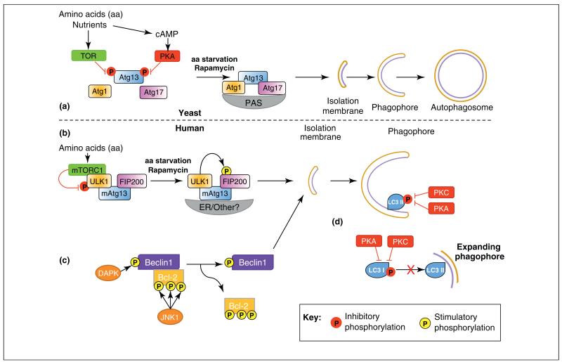
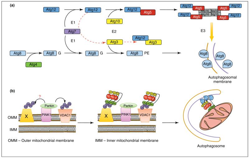
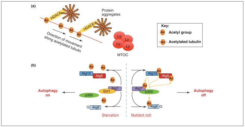
Autophagy is a highly conserved process that allows cells, tissues and organs to survive onslaughts such as nutrient deprivation, inflammation, hypoxia and other stresses. The core component proteins that regulate autophagy are well known, and the formation of a double-membrane structure that encompasses cytosolic cargo, including protein aggregates and organelles, has been intensively studied. However, less is known about the inputs that specifically alter recruitment of these components and how post-translational modifications can influence autophagy flux, or the rate at which autophagy substrates are turned over. We propose that three types of post-translational modifications - phosphorylation, ubiquitylation and acetylation - are crucial for autophagy induction, regulation and fine-tuning, and are influenced by a variety of stimuli. Understanding these novel mechanisms of autophagy regulation will give us deeper insights into this process and potentially open up therapeutic avenues.


Figures

Figure 1
Figure 1
Regulation of autophagy by phosphorylation. (a) Under nutrient-rich conditions in yeast, autophagy is inhibited by phosphorylation of Atg13 by target of rapamycin (TOR), preventing Atg1–Atg13–Atg17 complex formation. Phosphorylation of Atg13 by PKA inhibits Atg1–Atg13–Atg17 association with the pre-autophagosomal structure (PAS). Upon amino acid starvation or rapamycin treatment these inhibitory phosphorylations are removed, Atg1–Atg13–Atg17 complex forms and autophagy can proceed with isolation membrane, phagophore and eventually autophagosome formation. (b) In mammals, phosphorylation by mammalian-TOR (mTOR) complex 1 (mTORC1) of ULK1 inhibits ULK1 and initiation of autophagy. Amino acid starvation or rapamycin treatment causes dissociation of mTORC1 and ULK1 can phosphorylate and activate FIP200 allowing autophagy to proceed. (b) Phosphorylation of either Beclin1 or Bcl-2 by DAPK and JNK1, respectively, cause the dissociation of the Bcl-2/Beclin complex and is sufficient for autophagy induction and isolation membrane formation. (c) Phosphorylation of LC3 by either PKA or PKC is sufficient to inhibit LC3 incorporation into autophagosomes by an undefined mechanism. Hypothetical mechanisms include inhibition of the interaction of LC3 with LC3 interacting proteins or with the conjugation/lipidation machinery.
Figure 2
Figure 2
Regulation of autophagy by ubiquitylation. (a) The Ub-like proteins Atg8 (LC3/GABARAP in higher eukaryotes), Atg5 and Atg12 all undergo a Ub-like conjugation via the action of E1-(Atg7) and E2 (Atg10, Atg3)-like enzymes. Atg8 is primed by C-terminal cleavage by the protease Atg4, which exposes a C-terminal glycine and is then conjugated to PE. Atg12 is conjugated directly to Atg5 and forms a complex with Atg16 that possesses E3-like activity, driving Atg8-PE incorporation into expanding autophagosomes. An alternative conjugation is reported between the E2-like enzyme Atg3 and Atg12, which requires Atg7 (red broken line). (b) The E3-ligase Parkin is recruited to damaged mitochondria via the serine/threonine kinase PINK1 where it can ubiquitylate the target substrate VDAC1 (voltage dependent anion channel 1) with K27- and K63-linked polyUb chains. These chains serve to recruit p62/SQSTM1 and incorporate damaged mitochondria into autophagosomes. However, recent evidence suggests that VDAC1 is dispensable for Parkin mediated-mitochondrial clearance and that there might be other ubiquitylated proteins (Protein X) that recruit p62 (PB1, Phox and Bem1p domain; Znf, Zinc Finger domain; UBA, Ubiquitin-associated domain).
Figure 3
Figure 3
Regulation of autophagy by acetylation. (a) Adaptor proteins such as HDAC6 can interact with acetylated tubulin and transport protein aggregates along microtubules towards the microtubule organizing centre (MTOC) and the lysosomes (LY). (b) Under nutrient-rich conditions, the acetyltransferase p300 interacts with Atg7, acetylates the key autophagy proteins Atg7, Atg8, Atg12 and Atg5, and inhibits autophagy. During starvation, p300 dissociates and Sirt1 deacetylase exerts its activity, removing acetyl groups from Atg7, Atg5, Atg12 and Atg8 proteins, and allowing autophagy to proceed.

References

    1. Yang Z, Klionsky DJ. Eaten alive: a history of macroautophagy. Nat. Cell Biol. 2010;12:814–822. - PMC - PubMed
    1. Hayashi-Nishino M, et al. A subdomain of the endoplasmic reticulum forms a cradle for autophagosome formation. Nat. Cell Biol. 2009;11:1433–1437. - PubMed
    1. Yen WL, et al. The conserved oligomeric Golgi complex is involved in double-membrane vesicle formation during autophagy. J. Cell Biol. 2010;188:101–114. - PMC - PubMed
    1. van der Vaart A, et al. Exit from the golgi is required for the expansion of the autophagosomal phagophore in yeast Saccharomyces cerevisiae. Mol. Biol. Cell. 2010;21:2270–2284. - PMC - PubMed
    1. Hailey DW, et al. Mitochondria supply membranes for autophagosome biogenesis during starvation. Cell. 2010;141:656–667. - PMC - PubMed

Publication types