. 2011 Mar 31:178:196-207.
doi: 10.1016/j.neuroscience.2011.01.039. Epub 2011 Jan 26.

The majority of myelinated and unmyelinated sensory nerve fibers that innervate bone express the tropomyosin receptor kinase A

Affiliations

The majority of myelinated and unmyelinated sensory nerve fibers that innervate bone express the tropomyosin receptor kinase A

G Castañeda-Corral et al. Neuroscience. .

Abstract

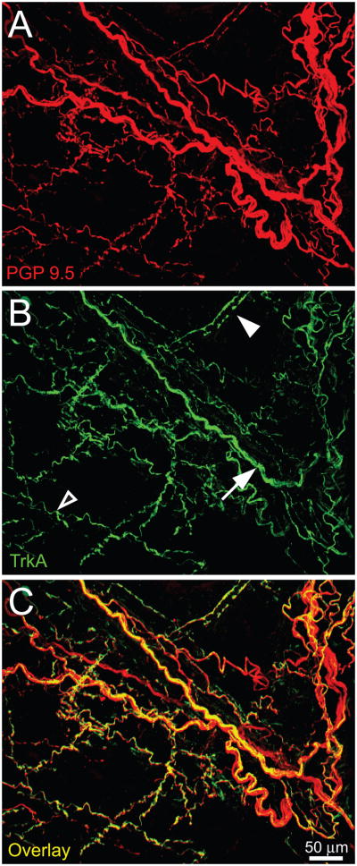
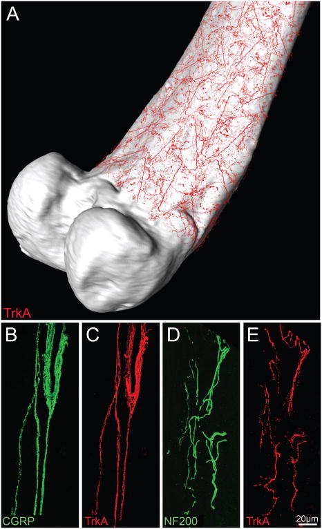
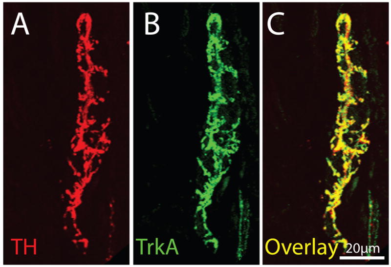
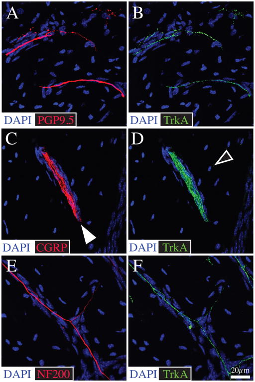
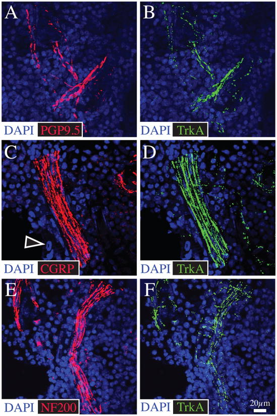
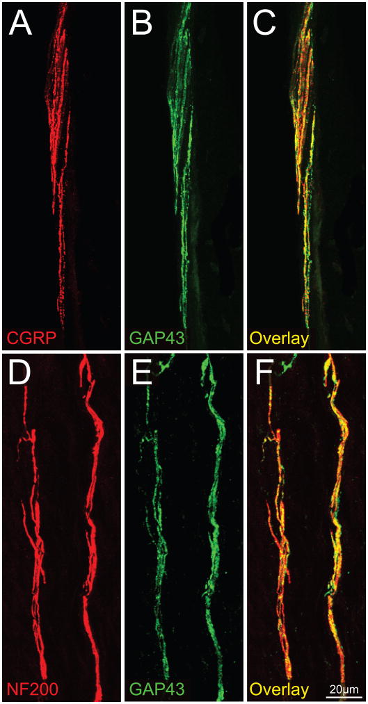
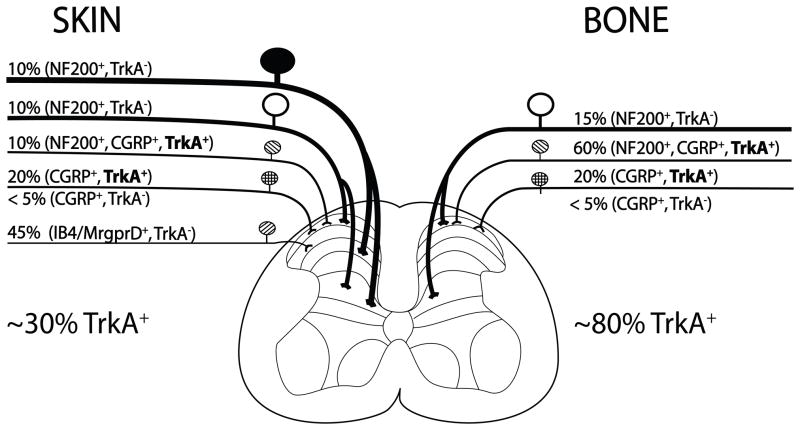
Although skeletal pain is a leading cause of chronic pain and disability, relatively little is known about the specific populations of nerve fibers that innervate the skeleton. Recent studies have reported that therapies blocking nerve growth factor (NGF) or its cognate receptor, tropomyosin receptor kinase A (TrkA) are efficacious in attenuating skeletal pain. A potential factor to consider when assessing the analgesic efficacy of targeting NGF-TrkA signaling in a pain state is the fraction of NGF-responsive TrkA+ nociceptors that innervate the tissue from which the pain is arising, as this innervation and the analgesic efficacy of targeting NGF-TrkA signaling may vary considerably from tissue to tissue. To explore this in the skeleton, tissue slices and whole mount preparations of the normal, adult mouse femur were analyzed using immunohistochemistry and confocal microscopy. Analysis of these preparations revealed that 80% of the unmyelinated/thinly myelinated sensory nerve fibers that express calcitonin gene-related peptide (CGRP) and innervate the periosteum, mineralized bone and bone marrow also express TrkA. Similarly, the majority of myelinated sensory nerve fibers that express neurofilament 200 kDa (NF200) which innervate the periosteum, mineralized bone and bone marrow also co-express TrkA. In the normal femur, the relative density of CGRP+, NF200+ and TrkA+ sensory nerve fibers per unit volume is: periosteum>bone marrow>mineralized bone>cartilage with the respective relative densities being 100:2:0.1:0. The observation that the majority of sensory nerve fibers innervating the skeleton express TrkA+, may in part explain why therapies that block NGF/TrkA pathway are highly efficacious in attenuating skeletal pain.


Figures

Figure 1
Figure 1
The mouse femoral periosteum is densely innervated with tropomyosin receptor kinase A (TrkA) expressing nerve fibers. Confocal micrographs of whole mount preparations of the femoral periosteum of naïve mice that were immunohistochemically labeled with antibodies against protein gene product 9.5 (PGP 9.5, red in A) and with TrkA (green in B) and a merged image of (A) and (B) is presented in (C). PGP 9.5 is a pan-neuronal marker that is frequently used to label all nerve fibers that innervate a structure. In the periosteum or bone the majority of PGP9.5+ nerve fibers express TrkA (C). Note that some TrkA+ nerve fibers have a unique morphology corresponding to a specific type of nerve fiber: some are observed as single thin nerve fibers (empty arrowhead, probably a CGRP+ C-fiber), bundles of thick nerve fibers (arrow, probably NF200 nerve fibers) and some appeared to enwrap blood vessels (filled arrowhead, probably TH+ sympathetic nerve fibers). Confocal images (30-μm z-series) were projected from 120 optical sections acquired at 0.25-μm intervals.
Figure 2
Figure 2
There is a high density of TrkA+ nerve fibers in the peristeum, which is a thin fibrous and cellular sheath that covers all but the articulated surface of the femur. For illustration purposes, representative confocal images of TrkA+ nerve fibers (in red, A) which were obtained from periosteal whole mount preparations, were overlaid onto a three-dimensional μCT image of the femur using Amira software. Panels B–E are representative confocal images of the periosteum obtained from frozen bone sections (20-μm-thick) that were double-stained with antibodies against calcitonin gene-related peptide (CGRP, B)/TrkA (C); and neurofilament 200 kDa (NF200, D)/TrkA (E). CGRP is a neuropeptide found predominantly in unmyelinated (C-fibers) and some myelinated sensory nerve fibers, whereas NF200 is expressed predominantly by myelinated primary afferent sensory nerve fibers (McCarthy and Lawson, 1990; Lawson and Wandell, 1991). Note that the majority of CGRP+ and NF200+ nerve fibers that innervate the periosteum also express TrkA. Images in B–C and D–E are from the same bone section. Confocal images of periosteum were acquired at 0.5 μm z-plane intervals and the total z-plane was 20 μm.
Figure 3
Figure 3
The majority of TH+ sympathetic nerve fibers that innervate the bone have a unique corkscrew appearance (as they wrap around blood vessels) and co-express TrkA. This figure contains confocal photomicrographs of periosteal frozen sections double-labeled with TH (A)/TrkA (B) and (C) is an overlay of (A) and (B). In mineralized bone and marrow one observes a similar co-localization in that nearly all TH+ nerve fibers are TrkA+. Images of periosteum were acquired from a frozen bone section (20-μm-thick) at 0.5 μm z-plane intervals and the total z-plane was 20 μm.
Figure 4
Figure 4
CGRP+ and NF200+ sensory nerve fibers in bone primarily travel in the Haversian and Volkmann canals and great majority of these nerve fibers co-express TrkA. Representative confocal images of bone frozen sections (20-μm-thick) double stained with antibodies against PGP 9.5 (A)/TrkA (B), CGRP (C)/TrkA (D), and NF200 (E)/TrkA (F). In the mineralized bone, CGRP+ and NF200+ fibers are typically thin, have a linear appearance and sometimes form nerve bundles. These nerve fibers are generally associated with blood vessels located in the Haversian canals (filled arrowhead). The bone sections were counterstained with DAPI (a nuclear marker) that labels nuclei of resident osteocytes which are abundant in mineralized bone. Confocal images of mineralized bone were acquired at 0.5 μm z-plane intervals and the total z-plane was 20 μm.
Figure 5
Figure 5
Bone marrow is innervated by both CGRP+ and NF200+ sensory nerve fibers and the great majority of these nerve fibers in bone marrow express TrkA. Representative confocal images obtained from frozen sections of bone (20 μm-thick) double-stained with antibodies against PGP 9.5 (A)/TrkA (B); CGRP (C)/TrkA (D); and NF200 (E)/TrkA (F). The cells which contain DAPI stained nuclei (blue) are primarily hematopoietic and stromal cells which comprise the marrow. The empty arrowhead in C points to the DAPI stained nucleus of a hematopoietic cell. Confocal images of the bone marrow were acquired at 0.5 μm z-plane intervals and the total z-plane was 20 μm.
Figure 6
Figure 6
Most NF200+ and CGRP+ sensory nerve fibers that innervate the bone also express growth associated protein 43 (GAP43). Confocal photomicrographs of periosteal frozen sections double labeled with CGRP (A)/GAP43 (B) and NF200 (D)/GAP43 (E). Panel (C) is an overlay of (A) and (B), while panel (F) is an overlay of (D) and (E). These data suggest that sensory nerve fibers that innervate the periosteum either are constantly remodeling and/or are capable of undergoing rapid sprouting and reorganization following bone injury or disease. Images of periosteum were acquired from a frozen bone sections (20-μm-thick) at 0.5 μm z-plane intervals and the total z-plane was 20 μm.
Figure 7
Figure 7
Schematic illustrating the approximate percentage (%) and types of sensory nerve fibers that innervate the skin vs. bone. The skin is innervated by thickly myelinated A-beta fibers (NF200+, TrkA), thinly myelinated A delta fibers (NF200+, TkA and NF200+, CGRP+, TrkA+), unmyelinated peptide-rich C fibers (CGRP+, TrkA+) and unmyelinated peptide-poor C-fibers (isolectin B4 (IB4)+), Mas related G protein-coupled receptor member D ((Mrgprd)+, TrkA). In contrast, the bone appears to be predominantly innervated by thinly myelinated A-delta fibers (NF-200+, TrkA and NF200+, CGRP+, TrkA+) and peptide-rich C-fibers (CGRP+ and TrkA+). In skin and bone there is also a small proportion (<5% of the total) of unmyelinated C-fibers that are CGRP+, TrkA. The percentages and types of sensory nerve fibers innervating the skin were estimated using data from previous studies (Bennett et al., 1996, Lu et al., 2001, Ambalavanar et al., 2005, Zylka et al., 2005) and for bone from the present and previous studies (Nakajima et al., 2008, Sugiura et al., 2008, Jimenez-Andrade et al., 2010b). Note that whereas approximately 30% of the sensory nerve fibers that innervate skin are TrkA+, greater than 80% of sensory nerve fibers that innervate the bone are TrkA+.

References

    1. Aguilar C, Vichinsky E, Neumayr L. Bone and joint disease in sickle cell disease. Hematol Oncol Clin North Am. 2005;19:929–941. viii. - PubMed
    1. Albrecht PJ, Hines S, Eisenberg E, Pud D, Finlay DR, Connolly MK, Pare M, Davar G, Rice FL. Pathologic alterations of cutaneous innervation and vasculature in affected limbs from patients with complex regional pain syndrome. Pain. 2006;120:244–266. - PubMed
    1. Ambalavanar R, Moritani M, Dessem D. Trigeminal P2X3 receptor expression differs from dorsal root ganglion and is modulated by deep tissue inflammation. Pain. 2005;117:280–291. - PubMed
    1. Andreev N, Dimitrieva N, Koltzenburg M, McMahon SB. Peripheral administration of nerve growth factor in the adult rat produces a thermal hyperalgesia that requires the presence of sympathetic post-ganglionic neurones. Pain. 1995;63:109–115. - PubMed
    1. Aoki Y, Ohtori S, Takahashi K, Ino H, Douya H, Ozawa T, Saito T, Moriya H. Expression and co-expression of VR1, CGRP, and IB4-binding glycoprotein in dorsal root ganglion neurons in rats: differences between the disc afferents and the cutaneous afferents. Spine. 2005;30:1496–1500. - PubMed

Publication types

MeSH terms