. 2011 Mar 1;186(5):3206-14.
doi: 10.4049/jimmunol.1003221. Epub 2011 Jan 28.

CX3CR1+ lung mononuclear phagocytes spatially confined to the interstitium produce TNF-α and IL-6 and promote cigarette smoke-induced emphysema

Affiliations

CX3CR1+ lung mononuclear phagocytes spatially confined to the interstitium produce TNF-α and IL-6 and promote cigarette smoke-induced emphysema

Zeyu Xiong et al. J Immunol. .

Abstract

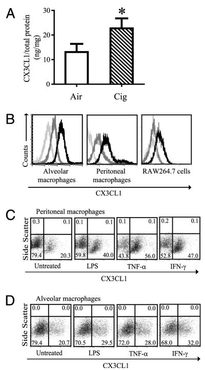
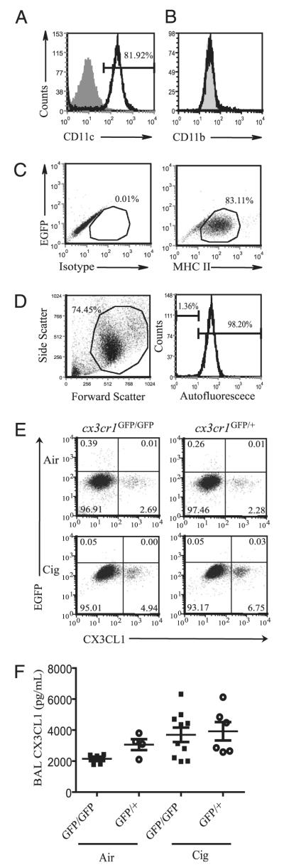
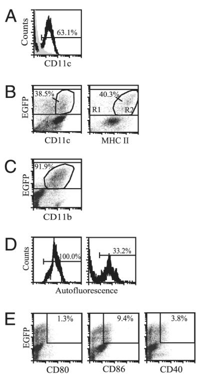
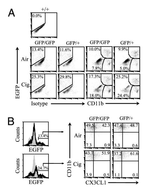
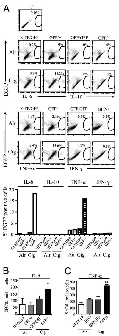
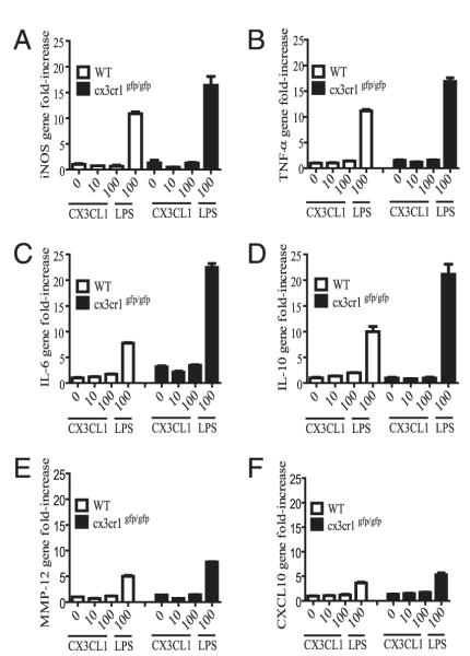
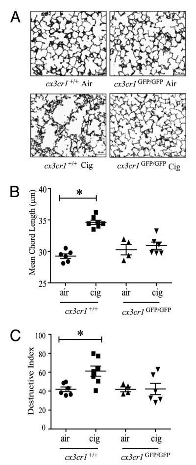
Increased numbers of macrophages are found in the lungs of smokers and those with chronic obstructive pulmonary disease. Experimental evidence shows the central role of macrophages in elaboration of inflammatory mediators such as TNF-α and the progression toward cigarette smoke-induced emphysema. We investigated the role of CX3CR1 in recruitment of mononuclear phagocytes, inflammatory cytokine responses, and tissue destruction in the lungs after cigarette smoke exposure. Using mice in which egfp is expressed at the locus of the cx3cr1 gene, we show that alveolar macrophages increased transmembrane ligand CX3CL1 expression and soluble CX3CL1 was detectable in the airspaces, but cx3cr1(GFP/GFP) and cx3cr1(GFP/+) mice failed to show recruitment of CX3CR1(+) cells into the airspaces with cigarette smoke. In contrast, cigarette smoke increased the accumulation of CX3CR1(+)CD11b(+) mononuclear phagocytes that were spatially confined to the lung interstitium and heterogenous in their expression of CD11c, MHC class II, and autofluorescent property. Although an intact CX3CL1-CX3CR1 pathway amplified the percentage of CX3CR1(+)CD11b(+) mononuclear phagocytes in the lungs, it was not essential for recruitment. Rather, functional CX3CR1 was required for a subset of tissue-bound mononuclear phagocytes to produce TNF-α and IL-6 in response to cigarette smoke, and the absence of functional CX3CR1 protected mice from developing tissue-destructive emphysema. Thus, CX3CR1(+) "tissue resident" mononuclear phagocytes initiate an innate immune response to cigarette smoke by producing TNF-α and IL-6 and are capable of promoting emphysema.


Figures

FIGURE 1
FIGURE 1
Transmembrane CX3CL1 expression on macrophages. A, Lung homogenates were obtained from C57BL/6 mice after air or cigarette smoke exposure for 8 wk and assayed for CX3CL1 expression. CX3CL1 concentration was normalized to the amount of total protein measured in each sample and is expressed as ng/mg ± SD. n = 8 mice per group. *p < 0.05. B, Mouse alveolar macrophages (left panel), peritoneal macrophages (middle panel), and mouse macrophage cell line RAW 264.7 cells (right panel) constitutively express transmembrane CX3CL1. Light-gray histogram, absence of immunostaining. Dark-gray histogram, isotype control staining. Black histogram, CX3CL1 staining. Representative images from four independent experiments are shown. C and D, Transmembrane CX3CL1 expression on peritoneal macrophages (C) and alveolar macrophages (D) is induced by TNF-α (20 ng/ml), IFN-γ (500 U/ml), and LPS (100 ng/ml) after 6 h stimulation (pooled cells from n = 9 to 12 mice).
FIGURE 2
FIGURE 2
Characterization of alveolar macrophages exposed to air or cigarette smoke. A and B, Alveolar macrophages express CD11c but not CD11b. Gray-filled histogram represents isotype control staining. Black histogram represents CD11c or CD11b staining. C, The majority of alveolar macrophages lack CX3CR1 and are MHC IIlow. Isotype control staining (left panel) versus MHC II staining (right panel). The y-axis indicates EGFP signal. D, BAL macrophages are autofluorescent high. BAL cells (left panel) with histogram of gated cell population shown (right panel). Autofluorescence shown was examined in the FL-2 channel in the absence of Ab staining. Representative data shown are from n = 2 to 3 cx3cr1GFP/+ mice per group, with two independent experiments conducted (A–D). E, Alveolar macrophages isolated from cx3cr1GFP/GFP and cx3cr1GFP/+ mice exposed to ambient air or cigarette smoke for 4 wk were assayed for CX3CR1 and transmembrane CX3CL1 expression (pooled samples from n = 3 mice per group). Representative data shown are from three independent experiments. F, Cleaved CX3CL1 concentrations in the BAL fluid of cx3cr1GFP/GFP and cx3cr1GFP/+ mice, air versus cigarette smoke exposure. Each point shown represents an individual mouse sample collected after 2- to 4-wk exposure, and data are expressed as mean ± SEM. p = 0.1 (ANOVA).
FIGURE 3
FIGURE 3
Characterization of interstitial lung mononuclear phagocytes after CD11c enrichment. A, The percentage of CD11c+ cells recovered from lung tissue digests after CD11c enrichment by magnetic bead positive selection. B, The percentage of CX3CR1+ lung mononuclear phagocytes, indicated by EGFP fluorescence, that express either CD11c (left panel) or MHC II (right panel). The percentages indicate a subset of total EGFP+ cells. C, The proportion of CX3CR1+ lung mononuclear phagocytes that express CD11b. D, The percentage of interstitial CX3CR1+ cells that are autofluorescent shows wide variability across samples, ranging from 20 to >90% (left and right panels are representative data from five independent experiments). E, The expression of costimulatory molecules CD80, CD86, and CD40 by CX3CR1+ cells is indicated. Cells were obtained from either cx3cr1GFP/GFP or cx3cr1GFP/+ mice under basal conditions as no significant differences in expression levels of surface markers were noted between cx3cr1GFP/GFP and cx3cr1GFP/+ mice. n = 2 to 3 mice from at least two independent experiments.
FIGURE 4
FIGURE 4
Accumulation of CX3CR1+CD11b+ mononuclear phagocytes in the lungs exposed to air or cigarette smoke. A, cx3cr1GFP/GFP and cx3cr1GFP/+ mice were exposed to ambient air or cigarette smoke for 4 wk, and CD11c-enriched interstitial cells were assayed for CD11b expression. The y-axis represents CX3CR1-expressing cells, indicated by EGFP fluorescence shift. The x-axis represents either isotype control staining or CD11b expression. EGFP fluorescence was gauged using cx3cr1+/+ mice cells set as negative control. Representative data shown from pooled cells from n = 3 mice per group, exposure time = 4 wk, two independent experiments conducted. B, The percentages of CX3CR1+ interstitial lung mononuclear phagocytes that express CD11b and transmembrane CX3CL1 after air versus cigarette smoke exposure are shown.
FIGURE 5
FIGURE 5
IL-6 and TNF-α responses in interstitial lung mononuclear phagocytes of cx3cr1GFP/GFP and cx3cr1GFP/+ mice exposed to air or cigarette smoke. A, CD11c-selected interstitial lung cells were isolated from mice exposed to ambient air or cigarette smoke for 4 wk, and CX3CR1-expressing cells (indicated by EGFP fluorescence shift) were assayed by intracellular cytokine immunostaining (pooled samples from n = 3 mice per group). The y-axis shows percentage of total CX3CR1 cells expressing cytokines, as indicated by EGFP fluorescence shift. EGFP fluorescence was gauged using cx3cr1+/+ mice cells set as negative control. GFP/GFP indicates cx3cr1GFP/GFP mice. GFP/+ indicates cx3cr1GFP/+ mice. B, ELISPOT data showing the number of IL-6–producing cells isolated from CD11c-enriched lung interstitial mononuclear phagocytes in cx3cr1GFP/GFP and cx3cr1GFP/+ mice exposed to air or cigarette smoke for 10 wk (pooled cells obtained from n = 5 mice in each group, performed in triplicate). *p < 0.05 (comparing cx3cr1GFP/GFP and cx3cr1GFP/+ mice; ANOVA). SFC, spot-forming cells. C, The number of TNF-α–producing cells from CD11c-enriched lung interstitial mononuclear phagocytes in cx3cr1GFP/GFP and cx3cr1GFP/+ mice exposed to cigarette smoke as determined by ELISPOT. Pooled cells were obtained from n = 2 to 3 mice in each group, 2-wk time exposure, with 6 to 12 wells per group. Data are expressed as mean ± SEM. **p < 0.01 (comparing cx3cr1GFP/GFP and cx3cr1GFP/+ mice; ANOVA).
FIGURE 6
FIGURE 6
A–F, Gene expression in interstitial lung mononuclear phagocytes obtained from cx3cr1+/+ and cx3cr1GFP/GFP mice. CD11c-enriched interstitial lung mononuclear phagocytes were harvested and pooled from cx3cr1+/+ (WT; n = 12) and cx3cr1GFP/GFP (n = 9) mice under steady-state conditions. Cells were stimulated in the presence or absence of increasing concentrations of recombinant CX3CL1 (0, 10, 100 nM) or LPS (100 ng/ml). Inducible NO synthase (iNOS), TNF-α, IL-6, IL-10, CXCL10, and MMP-12 gene expression was assayed by real-time PCR using the ΔΔCt method and 18S as the endogenous control. Unstimulated WT lung mononuclear phagocytes served as the calibrator. The y-axis represents gene fold-increase above the calibrator. Each condition was assayed in triplicate.
FIGURE 7
FIGURE 7
Effects of long-term cigarette smoke exposure on airspace enlargement in the lungs of cx3cr1GFP/GFP mice and cx3cr1+/+ WT control mice. A, Representative histologic sections of lungs from cx3cr1GFP/GFP and cx3cr1+/+ mice exposed to 24 wk of air versus cigarette smoke (Gill’s modified staining, original magnification ×200). B, Quantitative morphometric assessment of airspace enlargement in lung sections obtained from air-exposed and cigarette smoke-exposed cx3cr1+/+ and cx3cr1GFP/GFP mice (mean CL in μm). *p < 0.01 (ANOVA multiple comparisons). C, Destructive index of lung sections from air-exposed and cigarette smoke-exposed cx3cr1+/+ and cx3cr1GFP/GFP mice. Each point indicates individual mouse lungs studied expressed as mean ± SEM. *p < 0.05 (ANOVA).

References

    1. Mannino DM, Buist AS. Global burden of COPD: risk factors, prevalence, and future trends. Lancet. 2007;370:765–773. - PubMed
    1. Rabe KF, Hurd S, Anzueto A, Barnes PJ, Buist SA, Calverley P, Fukuchi Y, Jenkins C, Rodriguez-Roisin R, van Weel C, Zielinski J, Global Initiative for Chronic Obstructive Lung Disease Global strategy for the diagnosis, management, and prevention of chronic obstructive pulmonary disease: GOLD executive summary. Am. J. Respir. Crit. Care Med. 2007;176:532–555. - PubMed
    1. Stockley RA, Mannino D, Barnes PJ. Burden and pathogenesis of chronic obstructive pulmonary disease. Proc. Am. Thorac. Soc. 2009;6:524–526. - PubMed
    1. Hautamaki RD, Kobayashi DK, Senior RM, Shapiro SD. Requirement for macrophage elastase for cigarette smoke-induced emphysema in mice. Science. 1997;277:2002–2004. - PubMed
    1. Churg A, Wang RD, Tai H, Wang X, Xie C, Dai J, Shapiro SD, Wright JL. Macrophage metalloelastase mediates acute cigarette smoke-induced inflammation via tumor necrosis factor-alpha release. Am. J. Respir. Crit. Care Med. 2003;167:1083–1089. - PubMed

Publication types

MeSH terms