. 2011 May;39(10):4235-48.
doi: 10.1093/nar/gkr033. Epub 2011 Jan 29.

The intracellular sRNA transcriptome of Listeria monocytogenes during growth in macrophages

Affiliations

The intracellular sRNA transcriptome of Listeria monocytogenes during growth in macrophages

Mobarak A Mraheil et al. Nucleic Acids Res. 2011 May.

Abstract

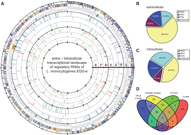
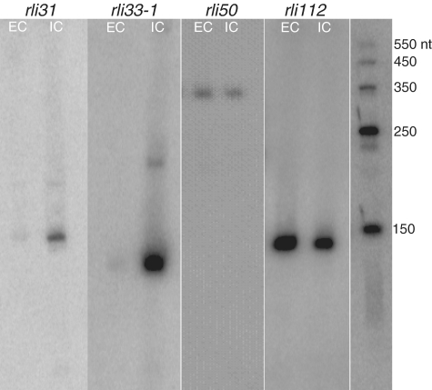
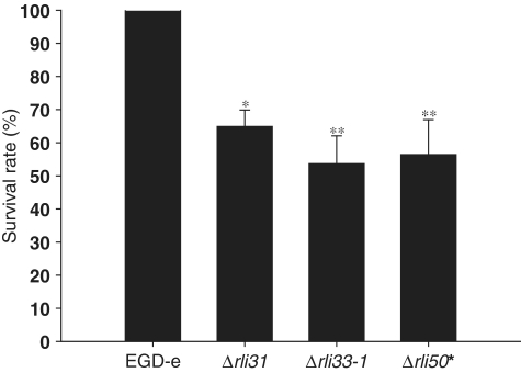
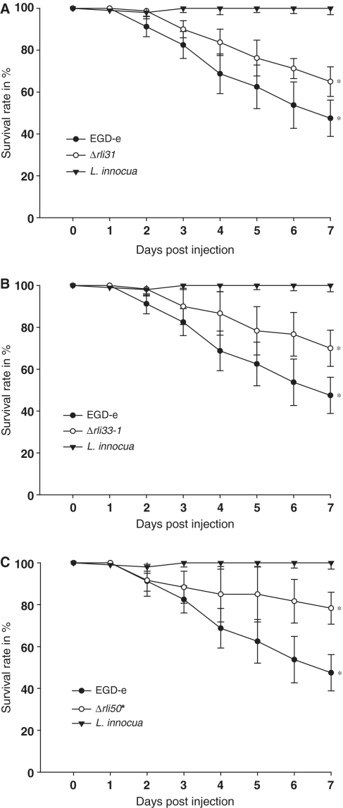
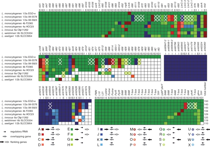
Small non-coding RNAs (sRNAs) are widespread effectors of post-transcriptional gene regulation in bacteria. Currently extensive information exists on the sRNAs of Listeria monocytogenes expressed during growth in extracellular environments. We used deep sequencing of cDNAs obtained from fractioned RNA (<500 nt) isolated from extracellularly growing bacteria and from L. monocytogenes infected macrophages to catalog the sRNA repertoire during intracellular bacterial growth. Here, we report on the discovery of 150 putative regulatory RNAs of which 71 have not been previously described. A total of 29 regulatory RNAs, including small non-coding antisense RNAs, are specifically expressed intracellularly. We validated highly expressed sRNAs by northern blotting and demonstrated by the construction and characterization of isogenic mutants of rli31, rli33-1 and rli50* for intracellular expressed sRNA candidates, that their expression is required for efficient growth of bacteria in macrophages. All three mutants were attenuated when assessed for growth in mouse and insect models of infection. Comparative genomic analysis revealed the presence of lineage specific sRNA candidates and the absence of sRNA loci in genomes of naturally occurring infection-attenuated bacteria, with additional loss in non-pathogenic listerial genomes. Our analyses reveal extensive sRNA expression as an important feature of bacterial regulation during intracellular growth.


Figures

Figure 1.
Figure 1.
Discovery of the intracellular sRNome of L. monocytogenes using RNA-Seq. (A) Extracellular and intracellular transcriptional landscape of L. monocytogenes is represented using GenomeViz (46). Circles display following information from outside to inside: (1) COG categories; (2) rRNAs and tRNAs (blue), a prophage-like locus (light brown) and the virulence gene cluster (red); (3) intracellular regulatory RNAs (outer circle) and extracellular regulatory RNAs (inner circle); (4) intracellular asRNAs (outer circle) and extracellular asRNAs (inner circle); (5) intracellular cis-regulatory RNAs including riboswitches (outer circle) and extracellular cis-regulatory RNAs including riboswitches (inner circle); (6) regulation of intracellular sRNAs; (7) regulation of intracellular asRNAs and (8) intracellular cis-regulatory RNAs including riboswitches; (B and C) Distribution of mapped sequence reads used for extracellular and intracellular transcriptome analysis; (D) Comparative analysis of sRNA transcriptome data using ‘cumulative’ values which can be summarized since sRNA candidates would not be counted multiple times (see Supplementary Figure S1 for a ‘non-cumulative’ version). Comparison of our RNA-seq results, whole genome tiling array from Toledo-Arana and coworkers (31), RNA-seq data of L. monocytogenes 10403S (32) and in silico regulatory RNA predictions (1).
Figure 2.
Figure 2.
Northern blots of sRNA candidates. Validation of RNA-seq data with northern blot analysis of extracellularly expressed sRNAs from bacteria at mid exponential growth phase in BHI (EC) compared with intracellularly expressed sRNAs at 4 h post-infection in murine macrophages (IC) of rli31, rli33-1, rli50 and rli112.
Figure 3.
Figure 3.
Survival of L. monocytogenes sRNA mutants in P388D1 murine macrophage cells. The macrophages were infected with the wild-type L. monocytogenes EGD-e and its isogenic deletion mutants, rli31, rli33-1 and rli50*, with an MOI of 10 in 24-well plates and bacterial CFU counts were measured on agar plated following lysis of the P388D1 cells after 4 h post-infection. n = 5; error bars indicate standard deviations (*P ≤ 0.005, **P ≤ 0.05).
Figure 4.
Figure 4.
Survival of Galleria mellonella larvae after inoculation with different L. monocytogenes sRNA mutants and L. innocua. Time course of survival of the larvae varies with the type of sRNA mutants employed for inoculation. Inoculation with 106 CFU/larvae EGD-e resulted in significantly higher killing rate of larvae in comparison to (A) rli31, (B) rli33-1 and (C) rli50*. The non-pathogenic L. innocua showed no mortality. Values represent means of at least three independent experiments ± standard deviations for 20 larvae per treatment (*P ≤ 0.005).
Figure 5.
Figure 5.
Mice infection studies with sRNA deletion mutants and of L. monocytogenes. Bacterial load in mice organs were also determined following in vitro infection with 2000 CFU of L. monocytogenes EGD-e wild-type strain as well as its isogenic sRNA mutants rli31, rli33-1 and rli50*. On day 3 after infection, the numbers of viable bacteria in spleens (A) and livers (B) of three animals per group were determined of wild-type EGD-e versus rli31, rli33-1 and rli50* in spleen and liver, respectively (n = 4). Error bars indicate standard deviations (*P ≤ 0.005, **P < 0.05).
Figure 6.
Figure 6.
Comparative overview of known and putative regulatory RNAs of L. monocytogenes EGD-e. EGD-e was compared with 3 L. monocytogenes serotypes (3 × 1/2a, 1 × 4b and 1 × 4a) and three non-pathogenic Listeria species (L. innocua, L. welshimeri and L. seeligeri). To determine the distribution of regulatory RNA inside the genus a BLAST analysis was conducted using sRNAdb (unpublished software). Candidates were considered present inside a strain in the case of a sequence identity of 60% and a coverage of 80%. Since the surrounding locus is often important for the function of the regulatory RNA, information about the conservation of adjacent genes was included using the same cutoff. Possible cases for direction, presence and absence of each regulatory RNA and its flanking genes was color-coded below. A white square indicates the absence of the regulatory RNA. As a reference for this analysis the relevant loci of L. monocytogenes EGD-e were chosen. The small black arrow depicted in the legend indicates the regulatory RNA, while larger arrows in black and white symbolize the left and right flanking gene, respectively. A large gray arrow denotes a gene overlapping the regulatory RNA in sense or antisense direction. The arrow direction is not representative for the strand but for the relation to the locus in the reference genome of L. monocytogenes EGD-e. (E) indicates extracellular and (I) intracellular expression of the regulatory RNA.

References

    1. Livny J, Teonadi H, Livny M, Waldor MK. High-throughput, kingdom-wide prediction and annotation of bacterial non-coding RNAs. PLoS ONE. 2008;3:e3197. - PMC - PubMed
    1. Storz G, Altuvia S, Wassarman KM. An abundance of RNA regulators. Annu. Rev. Biochem. 2005;74:199–217. - PubMed
    1. Vogel J, Sharma CM. How to find small non-coding RNAs in bacteria. Biol. Chem. 2005;386:1219–1238. - PubMed
    1. Gottesman S. Micros for microbes: non-coding regulatory RNAs in bacteria. Trends Genet. 2005;21:399–404. - PubMed
    1. Sorek R, Cossart P. Prokaryotic transcriptomics: a new view on regulation, physiology and pathogenicity. Nat. Rev. Genet. 2010;11:9–16. - PubMed

Publication types