. 2011 Mar 9;22(4):185-9.
doi: 10.1097/WNR.0b013e328344493c.

Loss of synaptic vesicles from neuromuscular junctions in aged MRF4-null mice

Affiliations

Loss of synaptic vesicles from neuromuscular junctions in aged MRF4-null mice

Qingbo Wang et al. Neuroreport. .

Abstract

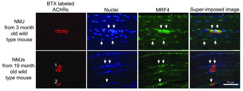
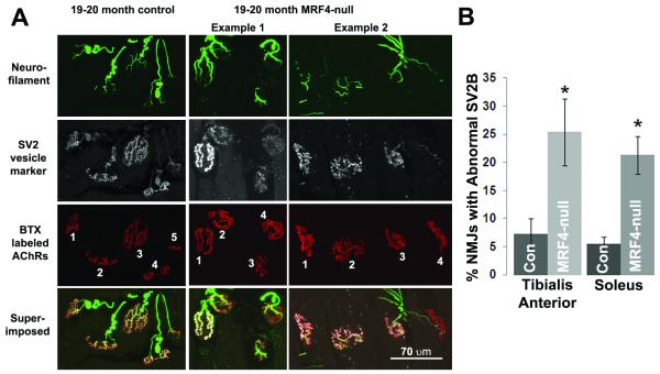
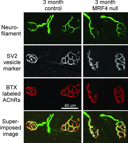
MRF4 belongs to the basic helix-loop-helix class of transcription factors and this and other members of its family profoundly influence skeletal muscle development. Less is known about the role of these factors in aging. As MRF4 is preferentially expressed in subsynaptic nuclei, we postulated it might play a role in maintenance of the neuromuscular junction. To test this hypothesis, we examined the junctional regions of 19-20-month-old mice and found decreased levels of SV2B, a marker of synaptic vesicles, in MRF4-null mice relative to controls. There was a corresponding decrease in grip strength in MRF4-null mice. Taken together, these data suggest that the intrinsic muscle factor, MRF4 plays an important role in maintenance of neuromuscular junctions.


Figures

Figure 1
Figure 1. MRF4 staining of neuromuscular junctions declines during aging
Shown are the staining of a representative neuromuscular junction (NMJ) from a young mouse (3 months old) and 2 NMJs from an old mouse (19 months old). NMJs are labeled by staining of acetylcholine receptors (AChRs) with α bungarotoxin (BTX). MRF4 staining in young muscle yielded a stereotyped pattern with a large region of very bright staining that encompassed the region containing multiple nuclei below the NMJ (downward arrows, upper row). In addition, MRF4 stained a subset of nuclei outside the NMJ (two examples, upward arrows, upper row). Two old NMJs are shown in the bottom row of images. While NMJ 2 had a region of bright MRF4 staining encompassing the NMJ (downward arrow near NMJ 2), no comparable region of MRF4 staining was present at NMJ1. Some nuclei near NMJ 1 were positive for MRF4 (downward arrows).
Figure 2
Figure 2. Absence of MRF4 worsens breakdown of NMJs during aging
A, Shown are 1 field of NMJs from an aged control mouse and 2 different fields from aged MRF4 null mice. Neurofilament staining of axons entering NMJs, SV2 staining of synaptic vesicles and α bungarotoxin (BTX) staining of acetylcholine receptors (AChRs) are all normal in this aged control mouse. NMJ 5 of this control mouse is on the edge and was not scored. In example 1 from a 20 month old MRF4 null muscle are 4 NMJs, all innervated by nerve as indicated by bright neurofilament staining. NMJ 1 has normal SV2 staining, but NMJs 2-4 have little SV2 staining, suggesting a marked reduction in the number of synaptic vesicles. In example 2 from a 19 month old mouse NMJs 1-3 have normal staining, but NMJ 4 has almost no SV2 staining. The neurofilament staining entering NMJ 4 consists of a thin line that is suggestive of a sprout that is reinnervating the NMJ (arrow). B, Quantification of the percentage of NMJs with “dim” SV2B staining was carried out in both aged control and MRF4-null muscles. A total of 20/269 NMJs in the control mice and 65/255 NMJs in the MRF4-null mice had reduced SV2B in TA muscles. To confirm that this phenomenon was not muscle type specific, three control and three MRF4-null soleus muscles were examined by the same approach with similar results. The asterisks indicate p < 0.05.
Figure 3
Figure 3. Aged Female MRF4-null Mice Have Reduced Grip Strength Relative to Controls
Forelimb grip strength was measured in aged (19-20 month old) female mice as described in the methods. The MRF4-null mice were significantly weaker in this test, with a mean grip strength of 1.95 ± 0.06 Newtons versus 2.3 ± 0.07 Newtons for the controls. Asterisks indicate p < 0.01. N = 14, control and n = 11, MRF4-null.
Figure 4
Figure 4. Absence of MRF4 has no discernable effect on NMJs from young animals
Shown are 1 field of NMJs from a young control mouse and 1 field from a young MRF4-null mouse. Neurofilament staining of axons entering NMJs, SV2 staining of synaptic vesicles and α bungarotoxin (BTX) staining of acetylcholine receptors (AChRs) are all normal.

References

    1. Deschenes MR. Effects of aging on muscle fibre type and size. Sports Med. 2004;34:809–24. - PubMed
    1. Vandervoort AA. Aging of the human neuromuscular system. Muscle Nerve. 2002;25:17–25. - PubMed
    1. Lang T, Streeper T, Cawthon P, Baldwin K, Taaffe DR, Harris TB. Sarcopenia: etiology, clinical consequences, intervention, and assessment. Osteoporos Int. 2010;21:543–559. - PMC - PubMed
    1. Luff AR. Age-associated changes in the innervation of muscle fibers and changes in the mechanical properties of motor units. Ann N Y Acad Sci. 1998;854:92–101. - PubMed
    1. Delbono O. Neural control of aging skeletal muscle. Aging Cell. 2003;2:21–9. - PubMed

Publication types

MeSH terms