. 2011 Mar;7(3):161-7.
doi: 10.1038/nchembio.521. Epub 2011 Jan 30.

Defining the geometry of the two-component proteasome degron

Affiliations

Defining the geometry of the two-component proteasome degron

Tomonao Inobe et al. Nat Chem Biol. 2011 Mar.

Abstract

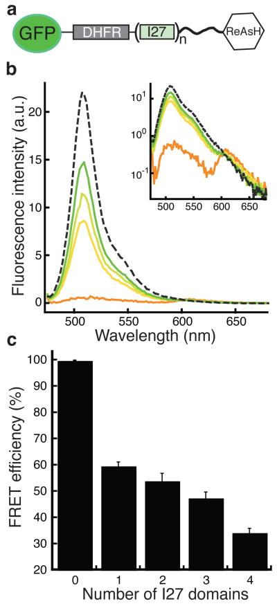
The eukaryotic 26S proteasome controls cellular processes by degrading specific regulatory proteins. Most proteins are targeted for degradation by a signal or degron that consists of two parts: a proteasome-binding tag, typically covalently attached polyubiquitin chains, and an unstructured region that serves as the initiation region for proteasomal proteolysis. Here we have characterized how the arrangement of the two degron parts in a protein affects degradation. We found that a substrate is degraded efficiently only when its initiation region is of a certain minimal length and is appropriately separated in space from the proteasome-binding tag. Regions that are located too close or too far from the proteasome-binding tag cannot access the proteasome and induce degradation. These spacing requirements are different for a polyubiquitin chain and a ubiquitin-like domain. Thus, the arrangement and location of the proteasome initiation region affect a protein's fate and are important in selecting proteins for proteasome-mediated degradation.


Figures

Figure 1
Figure 1
Proteasome-mediated degradation depends on initiation region length. (a) Linear representation of substrate proteins with initiation regions of different lengths. The substrates contained an E. coli DHFR domain and were targeted to the proteasome by an N-terminal Ub4 tag (left) or a UbL tag (right). Unstructured tails of different lengths were placed at the C-terminus of DHFR to serve as initiation regions. Degradation kinetics of substrates with ubiquitin (b) or UbL (c) tags and initiation regions of 15 (open triangles), 24 (black open squares), 29 (red solid diamonds), 34 (green solid circles), 44 (orange solid triangles), 64 (blue solid squares), and 102 (purple solid circles) amino acids derived from S. cerevisiae cytochrome b2. The extent of degradation was plotted as the percentage of protein remaining at different times. (d) Dependence of degradation rates (initial rates) on initiation region length for proteins with Ub4 (red solid diamonds) and UbL (green solid circles) tags. Data points represent mean values and error bars show standard errors calculated from three to five repeat experiments.
Figure 2
Figure 2
Separating the proteasome-binding tag and initiation region in degradation substrates. (a) Linear representation of proteasome substrates in which the proteasome-binding tag and initiation region are separated by the insertion of titin immunoglobulin I27 domains. The proteins contained a 44 amino acid long initiation region derived from cytochrome b2 at their C-termini and were targeted to the proteasome by either a Ub4 or a UbL tag at their N-termini. Structures of E. coli DHFR [PDB ID 1DRH] (b) and the immunoglobulin domain I27 of titin [PDB ID 1TIT] (c) shown in cartoon representation using PyMol (DeLano Scientific LLC, Palo Alto, CA; www.pymol.org).
Figure 3
Figure 3
Immunoglobulin domain I27 insertions increase the distance between proteasome-binding tag and the initiation region. In these experiments the tag at the N-terminus of DHFR was replaced by eGFP and the ReAsH-binding motif (CCGPCC) was fused to the C-terminus of the initiation region as shown in a schematic representation of these proteins (a). (b) Emission spectra of FRET substrates with no (orange), one (yellow), two (light green), or four (green) I27 domain insertions excited at 470 nm (4 nm bandwidth). After digestion with proteinase K, no FRET was observed (dashed line). Inset shows emission spectra with intensities shown on a log scale. (c) FRET efficiency for eGFP and ReAsH-labeled proteins decreased with the number of I27 domains inserted. Data shown by bar graph represent mean values and error bars show standard errors calculated from three repeat experiments.
Figure 4
Figure 4
Proteasome-mediated degradation depends on the spacing between proteasome-binding tag and initiation region. (a, b) Degradation kinetics for substrates with no (red solid diamonds), one (orange solid circles), two (black solid triangles), three (green solid squares), and four (blue solid square) I27 domain insertions targeted to the proteasome by either a Ub4 (a) or a UbL tag (b). (c) Plots of initial degradation rates as a function of the separation of the proteasome-binding tag and initiation region induced by the insertion of I27 domains show different relationships for substrates with Ub4 (red solid diamonds) and UbL tags (green solid circles). For proteins with ubiquitin tags, degradation rates were highest at short separations and then decreased at larger separations; for proteins with UbL tags, degradation was slowest at short separations and then accelerated with greater separation before decreasing again at the greatest separations. Data points represent mean values and error bars show standard errors calculated from three to five repeat experiments.
Figure 5
Figure 5
Spacing requirements for the two degron parts as initiation region length varies. To determine how the spacing of the proteasome-binding tag and initiation region modulates the effect of initiation region length on degradation, we varied proteasome-binding tag - initiation region spacing and initiation region length systematically as shown for constructs with UbL tags (a), Ub4-tagged substrates were constructed analogously. (b) Degradation kinetics for substrates in which three I27 domains separate a Ub4 tag from a 44 amino acid (green squares) or a 102 amino acid (light blue squares) initiation region. (c,d) Results for the degradation of other proteins in the array of constructs are shown as plots of initial degradation rates against initiation region lengths for constructs with UbL (c) and Ub4 (d) tags and zero (red solid diamonds), two (black solid circles), or four (blue solid triangles) I27 domain insertions. Degradation rates increase with the length of the initiation region for both tags but the strength of the response depends on the spacing. (e,f) Plots of initial degradation rates as a function of the distance between the proteasome-binding tag and initiation region for substrates with UbL (e) and Ub4 (f) tags with 34 amino acid (green solid diamonds), 44 amino acid (orange solid circles), or 102 amino acid (purple solid triangles) initiation regions. The longest initiation regions dampen the effect of degron spacing on degradation rates. Data points represent mean values and error bars show standard errors calculated from three to five repeat experiments.
Figure 6
Figure 6
Schematic representation of the length and spacing requirement for the initiation region. Proteasome caps contain ubiquitin and UbL receptors and these occupy different locations in the caps as indicated schematically, however, the placement of these locations in the sketch is arbitrary. Shapes and colors indicated different proteins. Light grey: proteasome caps; dark grey: proteasome core; circular arrows: ATPase subunits; scissors: proteolytic sites; grey circles: ubiquitin; yellow circle: UbL domain; blue shapes: substrates; yellow shapes: UbL-UBA adaptor protein; grey circles: ubiquitin binding sites; grey dotted circles: UbL binding sites. (a) Substrates in which both degron parts are close to each other are degraded efficiently when the receptors for ubiquitin tag and initiation region are close to each other on the proteasome. (b) When the ubiquitin tag and initiation region are farther apart in the substrate, the proteasome can no longer engage both degron parts at the same time and degradation is inefficient. (c) The spacing requirements for degrons with UbL tags may be explained if the UbL receptor on the proteasome is located at some distance from the initiation region receptor. (d) The figure shows a proteasome substrate bound to a UbL-UBA adaptor. In this representation the adaptor fits between the UbL and initiation site receptors on the proteasome. This spatial arrangement positions the substrate for effective degradation and keeps the unstructured regions on the adaptor away from the initiation region receptor.

References

    1. Pickart CM. Back to the future with ubiquitin. Cell. 2004;116:181–190. - PubMed
    1. Prakash S, Tian L, Ratliff KS, Lehotzky RE, Matouschek A. An unstructured initiation site is required for efficient proteasome-mediated degradation. Nat. Struct. Mol. Biol. 2004;11:830–837. - PubMed
    1. Takeuchi J, Chen H, Coffino P. Proteasome substrate degradation requires association plus extended peptide. EMBO J. 2007;26:123–131. - PMC - PubMed
    1. Schrader EK, Harstad KG, Matouschek A. Targeting proteins for degradation. Nat. Chem. Biol. 2009;5:815–822. - PMC - PubMed
    1. Thrower JS, Hoffman L, Rechsteiner M, Pickart CM. Recognition of the polyubiquitin proteolytic signal. EMBO J. 2000;19:94–102. - PMC - PubMed

Publication types

MeSH terms