. 2011 Mar;18(3):295-301.
doi: 10.1038/nsmb.1985. Epub 2011 Jan 30.

Single-molecule analysis of a molecular disassemblase reveals the mechanism of Hsc70-driven clathrin uncoating

Affiliations

Single-molecule analysis of a molecular disassemblase reveals the mechanism of Hsc70-driven clathrin uncoating

Till Böcking et al. Nat Struct Mol Biol. 2011 Mar.

Abstract

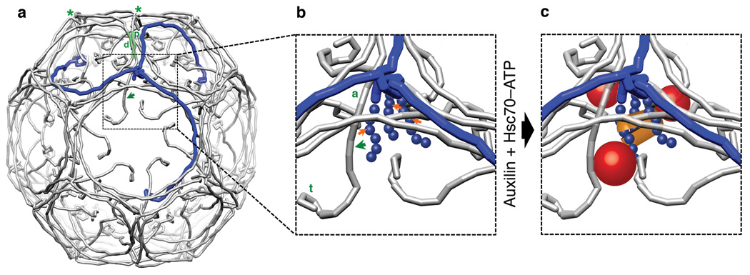
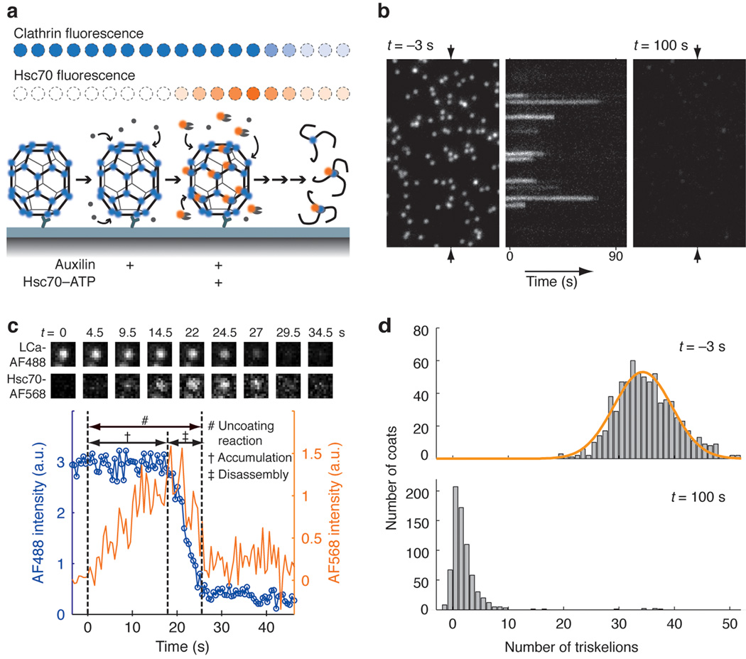
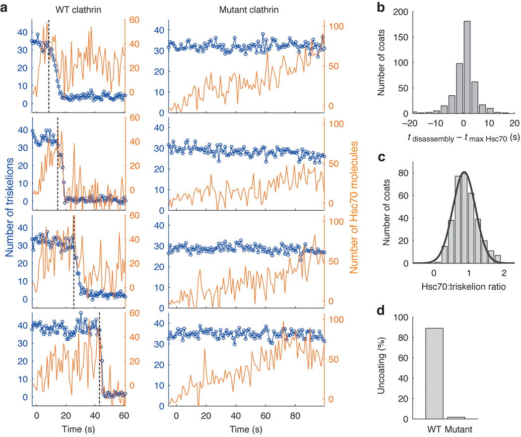
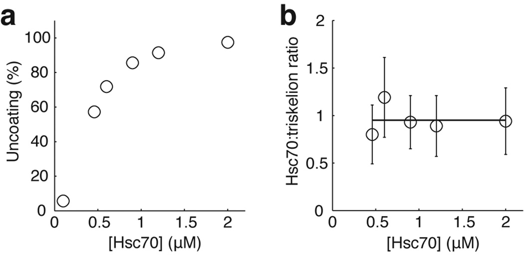
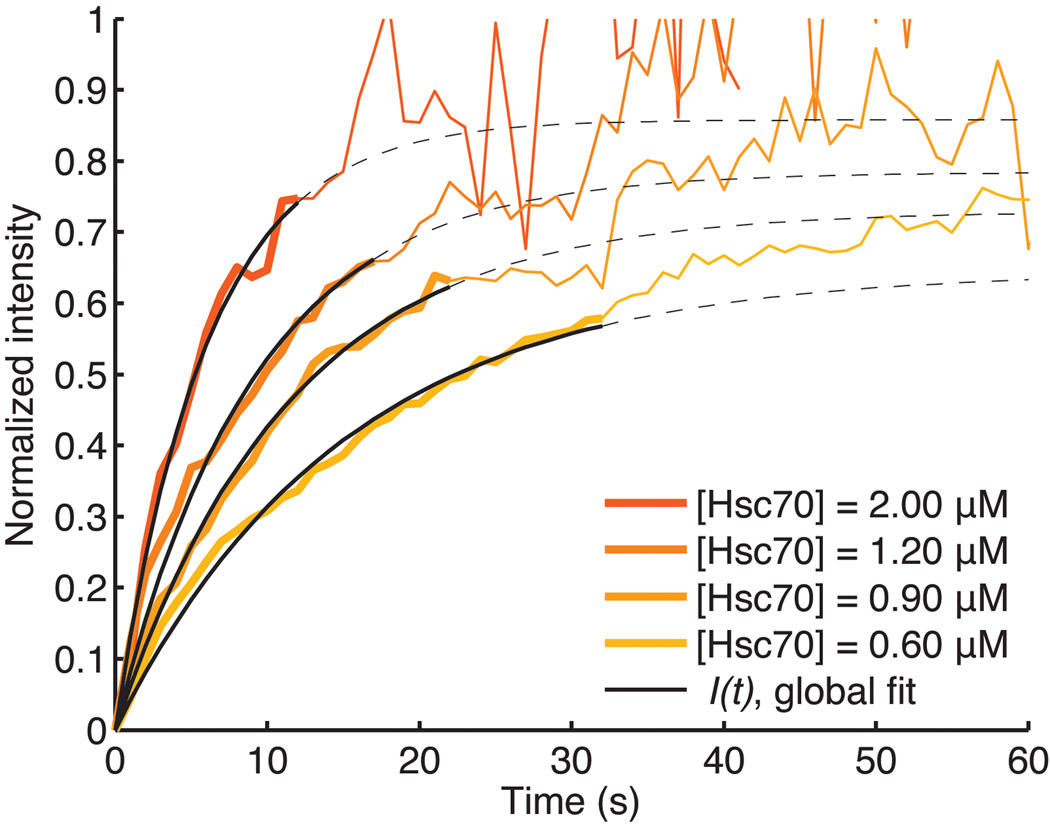
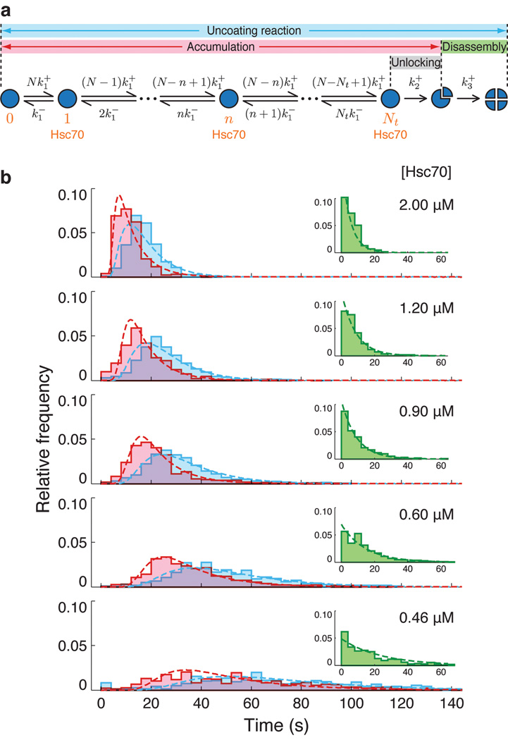
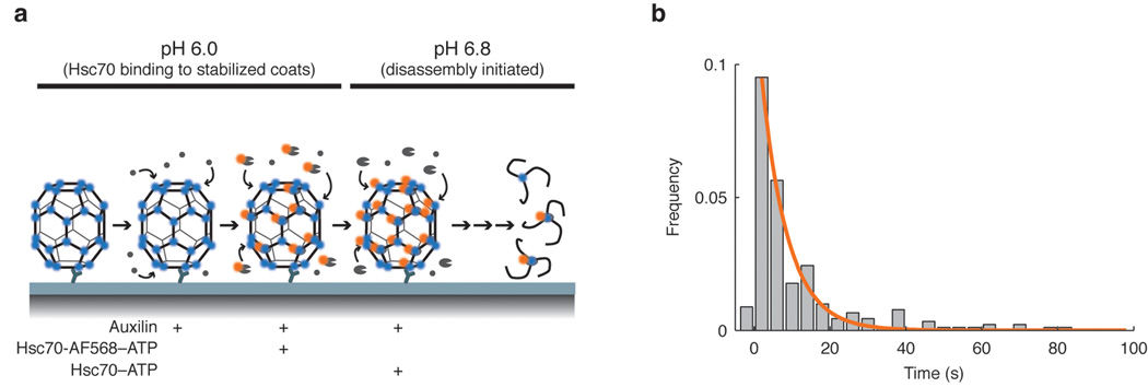
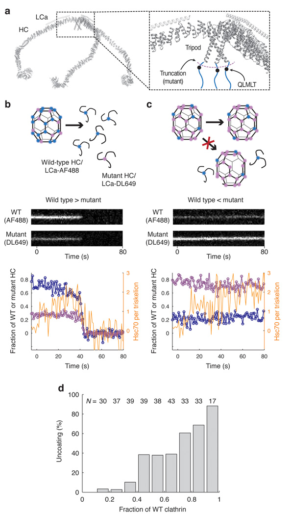
Heat shock cognate protein-70 (Hsc70) supports remodeling of protein complexes, such as disassembly of clathrin coats on endocytic coated vesicles. To understand how a simple ATP-driven molecular clamp catalyzes a large-scale disassembly reaction, we have used single-particle fluorescence imaging to track the dynamics of Hsc70 and its clathrin substrate in real time. Hsc70 accumulates to a critical level, determined by kinetic modeling to be one Hsc70 for every two functional attachment sites; rapid, all-or-none uncoating then ensues. We propose that Hsc70 traps conformational distortions, seen previously by cryo-EM, in the vicinity of each occupied site and that accumulation of local strains destabilizes the clathrin lattice. Capture of conformational fluctuations may be a general mechanism for chaperone-driven disassembly of protein complexes.


Figures

Figure 1
Figure 1. A clathrin coat with views of a vertex before and after formation of an uncoating intermediate
(a) Schematic representation of clathrin triskelions in a D6-barrel lattice (PDB 1XI4). One clathrin triskelion is highlighted in blue. The green shaded leg segments show the invariant contact between proximal (p) and distal (d) legs of the triskelions indicated by green asterisks at their hubs. The green arrow shows the direction of conformational shift when auxilin and Hsc70 bind. The hook-like elements at the (N-terminal) tips of the legs represent the β-propeller terminal domains. (b) Detail of a vertex before binding of auxilin and Hsc70. The unstructured C-termini of the clathrin heavy chain (blue balls), which contain the Q1638LMLT Hsc70-binding motif (orange arrows), extend inward from the helical tripod at the triskelion hub. The ankle (a) and terminal domain (t) shift in the direction of the green arrow when auxilin and Hsc70 bind. (c) Relative locations of bound auxilin (red spheres) and Hsc70 (orange lozenge) as determined by cryoEM ,. The shift in the positions of the clathrin ankle and terminal domain have been exaggerated to illustrate the expansion of the funnel surrounding the Hsc70-binding motif.
Figure 2
Figure 2. Single-particle visualization of clathrin uncoating
(a) Schematic representation of the single-particle uncoating assay. The intensities of fluorescence from labeled clathrin and Hsc70 were monitored by TIRF microscopy for clathrin/AP-2 coats captured onto the surface of a PEG modified glass coverslip. (b) Representative time series of a single-particle uncoating assay. The left and right panels correspond to the first and last frame of the fluorescence channel used to monitor the signal from coats tagged with clathrin LCa–AF488. The kymograph (middle) was generated from the vertical axis indicated by the arrows in the left panel. Hsc70–ATP (1.2 µM) arrived in the flow chamber at t = 0. It shows the unsynchronized disappearance of clathrin fluorescence. (c) Uncoating profile from a single coat. The selected snapshots from the time series (top) show the fluorescence from clathrin and Hsc70 in the selected coat, at various time points during the uncoating reaction carried out with 0.9 µM Hsc70. The snapshots are background-corrected averages of three successive frames. The plot shows intensity traces of the clathrin (blue) and Hsc70 (orange) signals. The t = 0 timepoint is the moment at which a rapid increase in Hsc70 background signal is recorded; this event corresponds to the arrival of Hsc70 within the evanescent field at the coverslip. (d) Histogram of the number of trimers (triskelions) per coat at the beginning (top) and the end (bottom) of the single particle uncoating assay carried out with 1.2 µM Hsc70. The number of trimers in intact coats follows a normal distribution with a mean of 34 triskelions per coat (top). In most cases, only one or two trimers remained at the site of a coat at the end of the reaction (bottom). Objects with overlapping point-spread functions were excluded from this analysis.
Figure 3
Figure 3. Requirement of the Hsc70 binding motif for Hsc70-driven uncoating
(a) Representative uncoating traces for clathrin/AP-2 coats assembled with wild-type heavy chain clathrin (left panels) or mutant heavy chain clathrin truncated at its C-terminus to remove the Hsc70 binding site (right panels). Hsc70–ATP (1.2 µM) arrived at t = 0 while monitoring the fluorescence intensities of clathrin (blue) and Hsc70 (orange) with TIRF illumination. Vertical dotted lines indicate onset of coat disassembly. The Hsc70:triskelion ratios at the onset of coat disassembly for the wild-type traces are 1.0, 0.8, 1.2 and 0.6 (from top to bottom). (b) Histogram of the time difference between maximal Hsc70 binding and onset of coat disassembly. (c) Histogram of the number of Hsc70 molecules bound per triskelion determined from the ratio of calibrated fluorescence intensities of Hsc70 to clathrin at the onset of coat disassembly (0.9±0.3, N = 350 coats). See Methods for details. (d) Uncoating efficiency for coats assembled with either wild type (N = 582 coats) or mutant clathrin (N = 660).
Figure 4
Figure 4. Hsc70 concentration dependence of the uncoating reaction
(a) Uncoating efficiency as a function of Hsc70 concentration. (b) Number of Hsc70 molecules bound per triskelion (mean ± s.d.) as a function of Hsc70 concentration determined from the ratio of the Hsc70 to the clathrin fluorescence intensity at the transition point between the accumulation and disassembly stages.
Figure 5
Figure 5. Recruitment of Hsc70 to clathrin/AP-2 coats during the accumulation phase follows first order kinetics
The orange traces correspond to the averaged intensity of the Hsc70 fluorescence measurements recorded in separate, single-particle uncoating assays with various Hsc70 concentrations. We used a global fit of the accumulation phase up to the mean accumulation time for each concentration (solid line) to determine the association and dissociation rates of Hsc70 binding. Mean accumulation times are derived from the accumulation time distributions in Figure 6. During this early recruitment phase Hsc70 binds preferentially to the QLMLT sequence after activation by the neighboring auxilin J-domain. During the later stages, when most of the specific sites have been occupied, activated Hsc70 is presumably delivered to other sites, which do not drive disassembly. The average Hsc70 binding curves become noisy beyond their corresponding mean accumulation times because most coats have started to disassemble, and the average is calculated from an increasingly lower number of coats. The dotted lines show the binding curves from the calculated parameters, beyond the range of the data used for fitting.
Figure 6
Figure 6. Kinetic model for the uncoating reaction
(a) Scheme for the kinetic analysis of Hsc70-driven uncoating. The model assumes that a threshold number of sites in the coat, Nt, must be occupied by a productively bound Hsc70 molecule to initiate disassembly. Hsc70 binds independently to the N binding positions in the coat (N = 36 for the D6 barrel), with a microscopic association rate constant k1+ (determined from the Hsc70 association curves, see Supplementary Methods and Fig. 5). The rate of any step is given by the product of k1+ and the number of unoccupied sites. Bound Hsc70 dissociates from the coat with an off-rate given by the microscopic dissociation rate constant, k1, and the number of bound Hsc70 molecules. Binding of Nt Hsc70 molecules triggers disassembly in a single, rate-limiting step of rate constant k2+ (determined independently, see Fig 7). The rate of clathrin dissociation, which depends linearly on Hsc70 concentration, is modeled with a single rate constant, k3+. (b) Distributions of the Hsc70 accumulation time (pink) and the full uncoating time (cyan), comprising accumulation and disassembly phases, at various Hsc70 concentrations, overlaid with fits of the kinetic model. The insets show disassembly time distributions (green) at various Hsc70 concentrations and the corresponding single exponential phase derived from the model fit.
Figure 7
Figure 7. Transition of Hsc70-loaded coats to the disassembly phase
(a) Experimental design used for the pH shift experiment. After immobilization of the clathrin/AP-2 coats onto the modified coverslip, the microfluidic chamber was perfused sequentially with auxilin, and then with a mixture of auxilin and Hsc70-AF568–ATP at pH 6. At this pH, the coats do not dissociate . To trigger uncoating, the pH was shifted to 6.8 in the presence of unlabeled Hsc70–ATP. Minimal uncoating was observed if the pH shift was done in the absence of Hsc70–ATP. (b) Histogram of the dwell times between pH shift and initiation of disassembly. The distribution of dwell times is a single exponential, indicating that the transition of an Hsc70-loaded coat to the disassembly phase has a single rate-limiting step, with rate constant k2+ (0.16 s−1).
Figure 8
Figure 8. Uncoating reaction with coats containing mixtures of wild type and mutant clathrin
(a) Backbone model of the heavy and light chains of a clathrin triskelion (PDB 3IYV) . The close-up of a triskelion hub shows the location of the binding site for Hsc70 (QLMLT motif, black dots) in the C- terminal unstructured region of the clathrin heavy chain (blue lines). (b, c) Outcome of the uncoating reaction for coats containing mostly wild type (b) or mostly mutant clathrin lacking the Hsc70-binding motif (c). Coats containing primarily wild type clathrin undergo normal disassembly with release of wild type and mutant clathrin (left); coats containing primarily mutant clathrin remain intact, and no wild type clathrin is released. Top, schematic representation of the coats and the outcome of the uncoating reaction. Middle, representative kymographs of single- particle uncoating traces from two coats, one containing excess wild-type clathrin (80%, left) and the other containing excess mutant clathrin (80%, right); wild-type and mutant clathrin were labeled with LCa–AF488 and LCa–DL649, respectively. Bottom, plots of fluorescence intensity traces; blue, wild type clathrin; purple, mutant clathrin; orange, Hsc70. (d) Uncoating efficiency as a function of the fraction of wild-type clathrin in the coat. Coats with different ratios of wild-type and mutant clathrin were prepared and mixed; their disassembly properties were then analyzed together in the same field (N = 311 coats).

References

    1. Dodson M, McMacken R, Echols H. Specialized nucleoprotein structures at the origin of replication of bacteriophage lambda. Protein association and disassociation reactions responsible for localized initiation of replication. J Biol Chem. 1989;264:10719–10725. - PubMed
    1. Hartl FU, Hayer-Hartl M. Converging concepts of protein folding in vitro and in vivo. Nat Struct Mol Biol. 2009;16:574–581. - PubMed
    1. Rapoport TA. Protein translocation across the eukaryotic endoplasmic reticulum and bacterial plasma membranes. Nature. 2007;450:663–669. - PubMed
    1. Mayer MP, Bukau B. Hsp70 chaperones: cellular functions and molecular mechanism. Cell Mol Life Sci. 2005;62:670–684. - PMC - PubMed
    1. Young JC, Barral JM, Ulrich Hartl F. More than folding: localized functions of cytosolic chaperones. Trends Biochem Sci. 2003;28:541–547. - PubMed

Publication types

MeSH terms