. 2011 Mar;18(3):288-94.
doi: 10.1038/nsmb.1978. Epub 2011 Jan 30.

Competing allosteric mechanisms modulate substrate binding in a dimeric enzyme

Affiliations

Competing allosteric mechanisms modulate substrate binding in a dimeric enzyme

Lee A Freiburger et al. Nat Struct Mol Biol. 2011 Mar.

Abstract

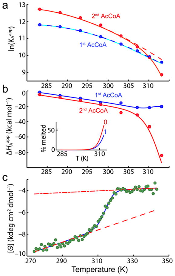
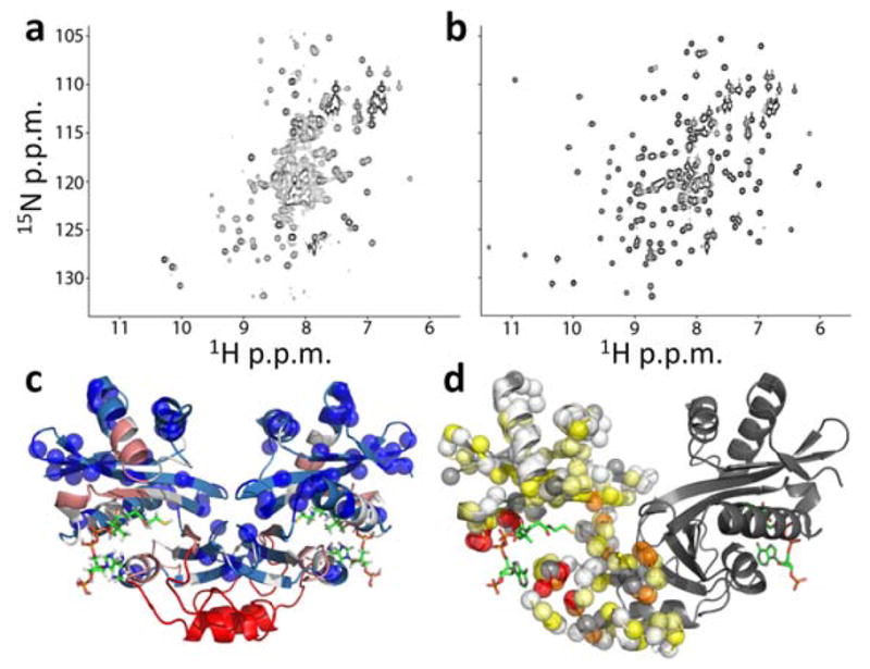
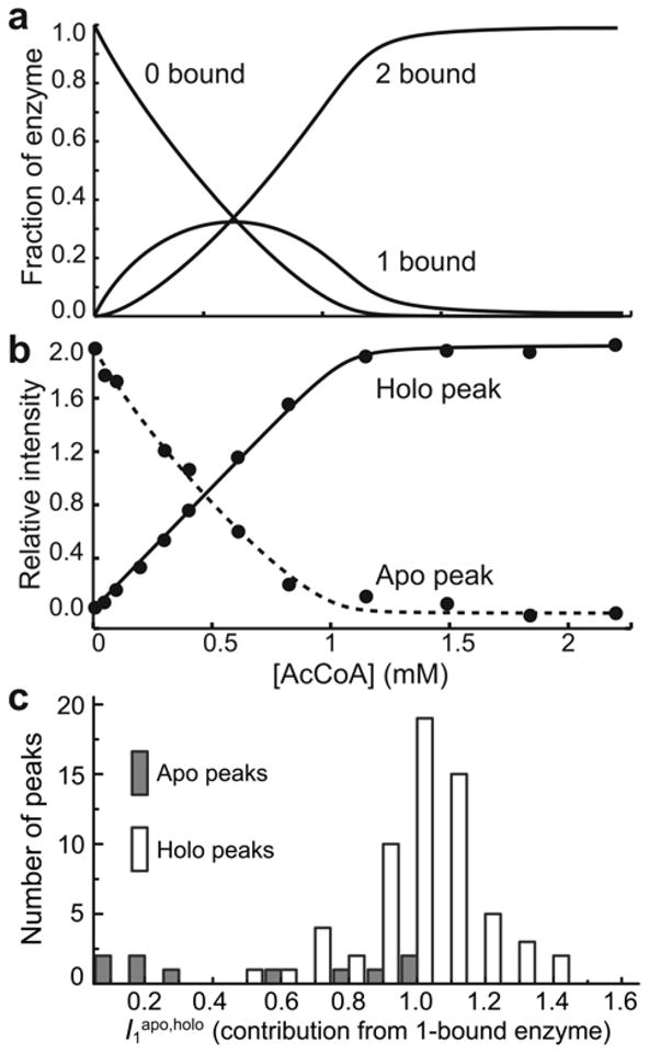
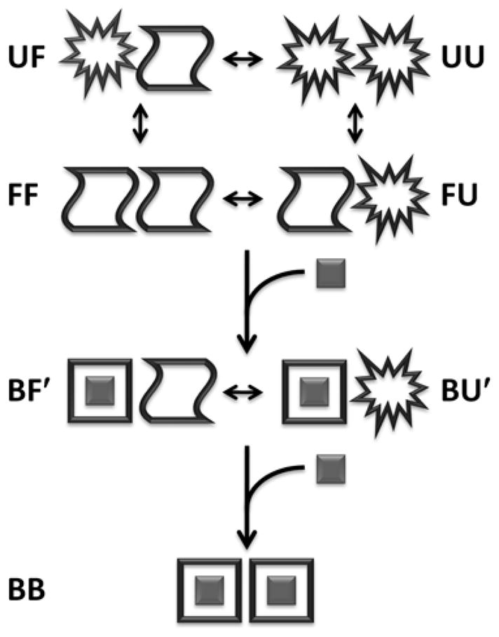
Allostery has been studied for many decades, yet it remains challenging to determine experimentally how it occurs at a molecular level. We have developed an approach combining isothermal titration calorimetry, circular dichroism and nuclear magnetic resonance spectroscopy to quantify allostery in terms of protein thermodynamics, structure and dynamics. This strategy was applied to study the interaction between aminoglycoside N-(6')-acetyltransferase-Ii and one of its substrates, acetyl coenzyme A. It was found that homotropic allostery between the two active sites of the homodimeric enzyme is modulated by opposing mechanisms. One follows a classical Koshland-Némethy-Filmer (KNF) paradigm, whereas the other follows a recently proposed mechanism in which partial unfolding of the subunits is coupled to ligand binding. Competition between folding, binding and conformational changes represents a new way to govern energetic communication between binding sites.


Figures

Figure 1
Figure 1
Schematic representation of homotropic allosteric models for a dimeric protein. ○ and □ correspond to subunits in binding-incompetent, and binding-competent states, respectively. (a) Monod-Wyman-Changeux (MWC): The symmetry of the dimer is preserved, so that only ○○ and □□ states are permitted. In the absence of ligand, both states are populated, while ligand binding forces the dimer into the □□ state. If the initial equilibrium favors the ○ ○ state, binding is positively cooperative, since the energetic cost of the ○ ○ to □ □ transition is paid by binding the first, but not the second ligand. Note that in the standard MWC model, both ○ ○ and □ □ bind ligand, but with different affinities. For the sake of simplicity we have shown the limiting case where ○ ○ is binding-incompetent. (b) Koshland-Nemethy-Filmer (KNF): Each subunit converts from the ○ to the ○ state only upon binding ligand. Cooperativity is explained in terms of the strengths subunit-subunit interactions. If the transition from the ○ ○ to ○ □ interface is energetically more favorable than from the ○ □ to □ □, binding is negatively cooperative, and the first ligand is bound more strongly than the second. If the transition from the ○ ○ to ○ □ interface is less favorable than from the ○ □ to □ □, binding is positively cooperative, and the second ligand is bound more strongly than the first. (c) Hilser-Thompson (HT): In this case, ○ and □ correspond to the unfolded and folded states, respectively. Each unbound subunit can populate either the folded or unfolded state, and the folding equilibrium is influenced by the state of the adjacent monomer. If folding (□) of one subunit promotes folding (□) of the adjacent subunit, binding is positively cooperative. Conversely, if folding (□) of one subunit promotes unfolding (○) of the adjacent subunit, binding is negatively cooperative.
Figure 2
Figure 2
Temperature dependence of AAC(6′)-Ii binding thermodynamics and secondary structure. (a) Equilibrium association constants for the first and second molecules of AcCoA, plotted as a function of temperature. Solid lines correspond to the best fit obtained with Eqs. 2 to 9. Dashed lines indicate the predicted affinities in the absence of thermal melting of the subunits. (b) Binding enthalpies of the first and second molecules of AcCoA. Inset shows the fraction of free subunits that are melted in the 0-bound and 1-bound forms. (c) Molar ellipticity (222 nm) of AAC(6′)-Ii as a function of temperature. Dashed and dash-dot lines correspond to the pre- and post-transition baselines, respectively.
Figure 3
Figure 3
Changes in AAC(6′)-Ii NMR spectra produced by AcCoA binding. (a,b) 1H/15N correlation spectra of AAC(6′)-Ii free (a) and saturated with AcCoA (b). (c) X-ray crystal structure of the enzyme (PDB 2A4N) bound to CoA (sticks) with blue spheres indicating the locations of residues with assigned cross-peaks in apo spectra. The backbone is color-coded according to the distance in the 1° sequence (n) from the nearest assigned residue according to 1≤n≤2 (light blue), 3≤n≤5 (white), 6≤n≤10 (pink), n>10 (red) (d) Apparent minimal chemical shift differences ( Δδapp=(10×Δδapp1H)2+(Δδapp15N)2) between the free and bound states, mapped onto the X-ray crystal structure (PDB 2A4N). Backbone amide nitrogen atoms are indicated with spheres for one subunit of the dimer and colored according to Δδapp<0.5 ppm (white), 0.5≤Δδapp<1 (light yellow), 1≤Δδapp<2 (yellow), 2≤Δδapp<4 (orange), 4≤Δδapp (red). Unassigned residues, including prolines, are indicated with gray spheres. Structures were generated using PyMOL.
Figure 4
Figure 4
Analysis of NMR titration data. (a) Fraction of the enzyme in the 0-bound, 1-bound, and 2-bound states determined by ITC. (b) Intensities of the apo (dashed line) and holo (solid line) peaks for Leu56 as a function of [AcCoA]. The intensities were analyzed to extract the relative contribution of the 1-bound enzyme to the signals (I1apo,holo) as described in the text. The lines correspond to the optimized theoretical intensities. (c) Histograms of the relative contribution of the 1-bound enzyme to apo (I1apo) and holo (I1holo) peaks in titrations of AAC(6′)-Ii with AcCoA.
Figure 5
Figure 5
Schematic representation of the allosteric binding model. B corresponds to the bound state. F(F′) and U(U′) correspond to the free folded and partially unfolded states that are adjacent to a free (bound) subunit, respectively. Transitions involving the symmetry-related FB and UB states are not shown.
Figure 6
Figure 6
Dependence of the apparent cooperativity coefficient (αapp) on subunit instability (KU0=[UF]/[FF]) calculated using Eq. [1] and (a) the intrinsic cooperativity coefficient (αint=1.3) and ratio of melting equilibrium constants constants (φ=1.9) determined for AAC(6′)-Ii at 37°C and (b) hypothetical proteins in which αint=φ=4,16,64,256,1024. The circle in (a) corresponds to the KU0 value obtained for AAC(6′)-Ii at 37°C (0.2).

References

    1. Perutz MF. Mechansism of cooperativity and allosteric regulation in proteins. Q Rev Biophys. 1989;22:139–236. - PubMed
    1. Monod J, Wyman J, Changeux JP. On nature of allosteric transitions - a plausible model. J Mol Biol. 1965;12:88–118. - PubMed
    1. Fenton AW. Allostery: an illustrated definition for the ‘second secret of life’. Trends Biochem Sci. 2008;33:420–425. - PMC - PubMed
    1. DeDecker BS. Allosteric drugs: thinking outside the active-site box. Chem Biol. 2000;7:R103–R107. - PubMed
    1. Koshland DE, Nemethy G, Filmer D. Comparison of experimental binding data and theoretical models in proteins containing subunits. Biochemistry. 1966;5:365–385. - PubMed

Publication types

Substances

Grants and funding