

doi: 10.1038/nsmb.1982.
Epub 2011 Jan 30.
Retinal dynamics underlie its switch from inverse agonist to agonist during rhodopsin activation
Affiliations
- PMID: 21278756
- PMCID: PMC5283944
- DOI: 10.1038/nsmb.1982
Item in Clipboard
Retinal dynamics underlie its switch from inverse agonist to agonist during rhodopsin activation
Nat Struct Mol Biol.
2011 Mar.
Abstract
X-ray and magnetic resonance approaches, though central to studies of G protein-coupled receptor (GPCR)-mediated signaling, cannot address GPCR protein dynamics or plasticity. Here we show that solid-state (2)H NMR relaxation elucidates picosecond-to-nanosecond-timescale motions of the retinal ligand that influence larger-scale functional dynamics of rhodopsin in membranes. We propose a multiscale activation mechanism whereby retinal initiates collective helix fluctuations in the meta I-meta II equilibrium on the microsecond-to-millisecond timescale.
Figures

Site-specific 2H NMR relaxation illuminates functional dynamics of retinylidene methyl groups within binding pocket of rhodopsin. (a) Light absorption yields 11-cis to trans isomerization, converting retinal from an inverse agonist to an agonist by a series of intermediates with different time scales. (b–c) Solid-state 2H NMR spectra for dark-state rhodopsin with 11-cis retinal deuterated at C5-, C9-, or C13–C2H3 groups in POPC bilayers (1:50 molar ratio). The 2H NMR lineshapes indicate rapid axial spinning of C–C2H3 groups down to at least −160 °C. (e,f) Partially relaxed 2H NMR spectra for retinylidene C9- and C13–C2H3 groups of rhodopsin in aligned POPC membranes (θ=0°) at −150 °C. (g) Inversion-recovery plots showing site-specific variations in spin-lattice (T1Z) relaxation times for C9- and C13-Me groups at −150 °C.
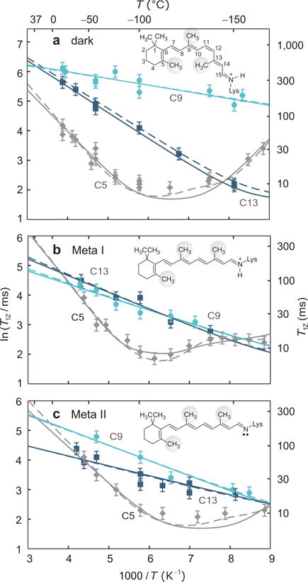
Solid-state 2H NMR of captures site-specific changes in retinal mobility during light activation of rhodopsin. Spin-lattice (T1Z) relaxation times (±s.d.) of retinylidene methyl groups are shown versus reciprocal temperature in (a) the dark, (b) Meta I, and (c) Meta II states (−30 to −160 °C). Methyl dynamics are described by an axial 3-fold jump model or a continuous diffusion model with coefficients D‖ and D⊥. In panels a–c rotation about the methyl 3-fold (C3) axis corresponds to solid lines with D⊥=0; the dashed lines include restricted off-axial diffusion (D⊥=D‖). Fits for the C5-Me in Meta I in panel b assume unlike rotational diffusion constants (D‖≠D⊥) (dashed line), or two conformers with different bond orientations and axial diffusion coefficients (solid line).

2H NMR relaxation of retinal sheds new light on activation mechanism of rhodopsin. (a) Summary of analysis of solid-state 2H NMR measurements. Order parameters of rapidly spinning methyl groups are designated by
; the pre-exponential factor is k0 for 3-fold axial jumps or D0 for continuous diffusion; and Ea indicates the activation energy. (The diffusion model assumes either D⊥=0 (right) or ηD ≡ D‖/D⊥=1 (left) except for the C5-Me in Meta I, where ηD≠1.) (b–d) Proposed activation mechanism for rhodopsin in membranes based on X-ray, FTIR, and 2H NMR data (see text). Isomerization of retinal displaces the E2 loop towards the extracellular (e) side with fluctuations of TM helices H5 and H6 exposing transducin (Gt) recognition sites on the opposing cytoplasmic (c) surface. Figure produced (PDB code 1U19) using PyMOL [http://pymol.sourceforge.net/ ]
References
-
- Kobilka B, Schertler GFX. Trends Pharmacol Sci. 2008;29:79–83. - PubMed
-
- Ahuja S, Smith SO. Trends Pharmacol Sci. 2009;30:494–502. - PubMed
-
- Okada T, et al. J Mol Biol. 2004;342:571–583. - PubMed
-
- Ridge KD, Palczewski K. J Biol Chem. 2007;282:9297–9301. - PubMed
-
- Brown MF. J Chem Phys. 1982;77:1576–1599.
Publication types
MeSH terms
Substances
Grants and funding
LinkOut - more resources
Full Text Sources
