

doi: 10.1016/j.jsmc.2010.08.003.
Neuropharmacology of Sleep and Wakefulness
Affiliations
- PMID: 21278831
- PMCID: PMC3026477
- DOI: 10.1016/j.jsmc.2010.08.003
Item in Clipboard
Neuropharmacology of Sleep and Wakefulness
Sleep Med Clin.
2010 Dec.
Abstract
The development of sedative/hypnotic molecules has been empiric rather than rational. The empiric approach has produced clinically useful drugs but for no drug is the mechanism of action completely understood. All available sedative/hypnotic medications have unwanted side effects and none of these medications creates a sleep architecture that is identical to the architecture of naturally occurring sleep. This chapter reviews recent advances in research aiming to elucidate the neurochemical mechanisms regulating sleep and wakefulness. One promise of rational drug design is that understanding the mechanisms of sedative/hypnotic action will significantly enhance drug safety and efficacy.
Figures
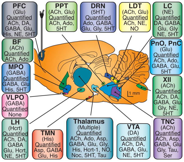
Sagittal drawing of the rat brain (modified from197) schematizes the location, shape, and size of some brain regions that regulate sleep and wakefulness. The name of each brain region appears in bold print, the major neurotransmitters used for signaling to other brain regions are in parentheses, and neurochemical analytes relevant for arousal-state control that have been measured in that brain region are listed under the header “Quantified ”. The microdialysis probe is drawn to scale and is shown sampling from the prefrontal cortex. Abbreviations: XII – hypoglossal nucleus; BF – basal forebrain; DRN – dorsal raphé nucleus; LC – locus coeruleus; LDT – laterodorsal tegmental nucleus; LH – lateral hypothalamus; MPO – medial preoptic area; PFC – prefrontal cortex; PPT – pedunculopontine tegmental nucleus; PnC – pontine reticular formation, caudal part; PnO – pontine reticular formation, oral part; TMN – tuberomamillary nucleus; TNC – trigeminal nucleus complex; VLPO – ventrolateral preoptic area; VTA – ventral tegmental area; 5HT – serotonin; ACh – acetylcholine; Ado – adenosine; Asp – aspartate; DA – dopamine; GABA – γ-aminobutyric acid; Glu – glutamate; Gly – glycine; His – histamine; Hcrt – hypocretin; NE – norepinephrine; NO – nitric oxide; Noc – nociceptin; Ser – serine; 5HT – serotonin; Tau – taurine. Figure reprinted from Watson et al., 2010 with permission.
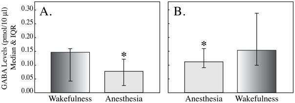
Consistent results were obtained when measuring GABA during the transition from wakefulness to anesthesia (A) and during the resumption of wakefulness after anesthesia (B). In both cases GABA was significantly decreased during the loss of wakefulness caused by isoflurane anesthesia. Data reprinted from Vanini et al., 2008 with permission.

Top: schematic coronal section of rat brain stem illustrates placement of a microdialysis probe in the PRF. Ringer’s solution is pumped into the probe and samples are collected for quantification of ACh. Schematized at top right of brain are electrodes and an amplifier for recording the cortical electroencephalogram (EEG), and a representative trace showing EEG activity after intravenous administration of eszopiclone. Bottom: Histograms summarize the significant decrease in ACh release within the PRF caused by intravenous administration of eszopiclone. Data reprinted from Hambrecht-Wiedbusch et al., 2010 with permission.
References
-
- Watson CJ, Baghdoyan HA, Lydic R. A neurochemical perspective on states of consciousness. In: Hudetz AG, Pearce RA, editors. Suppressing the Mind: Anesthetic Modulation of Memory and Consciousness. New York: Springer/Humana Press; 2010. pp. 33–80.
-
- Steriade M, McCarley RW, editors. Brain Control of Wakefulness and Sleep. New York: Kluwer Academic/Plenum Publishers; 2005.
-
- McCarley RW. Neurobiology of REM and NREM sleep. Sleep Med. 2007;8:302–330. - PubMed
Grants and funding
LinkOut - more resources
Full Text Sources
Other Literature Sources
