

Electrosurgical excision of acne keloidalis nuchae with secondary intention healing
- PMID: 21278897
- PMCID: PMC3030213
Electrosurgical excision of acne keloidalis nuchae with secondary intention healing
Abstract
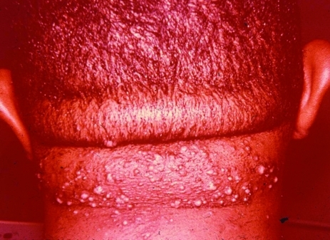
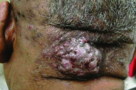
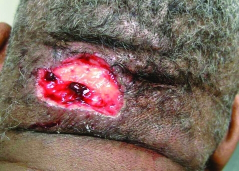
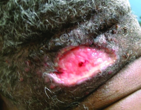
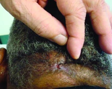
Acne keloidalis nuchae is an idiopathic, scarring folliculitis characterized by the formation of papules and pustules that may ultimately develop into tumor-like masses distributed on the nape of the neck and occipital region of the scalp. This hyperproliferative disorder is most commonly seen in African-American men. While the pathogenesis remains uncertain, precipitating factors include localized trauma, chronic irritation, seborrhea, and androgen excess. The treatment of acne keloidalis nuchae is challenging and depends on the clinical stage of the disease; however, a multifaceted approach involving combination therapies has proven to be effective in some cases. Excision with healing by secondary intention is a recommended option for patients with large plaque- and tumor-stage acne keloidalis nuchae. This case report reviews the management of a patient with tumor-stage acne keloidalis nuchae successfully treated with electrosurgical excision and secondary intention healing with excellent cosmetic results and no recurrence of the disease.
Figures
Similar articles
-
Electrosurgical excision and loop cautery of acne keloidalis nuchae: alternatives to standard surgical excision.J Cosmet Laser Ther. 2025;27(4-5):163-165. doi: 10.1080/14764172.2025.2510436. Epub 2025 May 23. J Cosmet Laser Ther. 2025. PMID: 40410935
-
Surgical excision of acne keloidalis nuchae with secondary intention healing.Clin Exp Dermatol. 2008 Jan;33(1):53-5. doi: 10.1111/j.1365-2230.2007.02549.x. Epub 2007 Oct 9. Clin Exp Dermatol. 2008. PMID: 17927781
-
Acne Keloidalis Nuchae: A Staged Reconstruction.Cureus. 2021 Sep 21;13(9):e18173. doi: 10.7759/cureus.18173. eCollection 2021 Sep. Cureus. 2021. PMID: 34692352 Free PMC article.
-
Acne keloidalis in females: case report and review of literature.J Natl Med Assoc. 2005 May;97(5):736-8. J Natl Med Assoc. 2005. PMID: 15926654 Free PMC article. Review.
-
Acne keloidalis nuchae: prevalence, impact, and management challenges.Clin Cosmet Investig Dermatol. 2016 Dec 14;9:483-489. doi: 10.2147/CCID.S99225. eCollection 2016. Clin Cosmet Investig Dermatol. 2016. PMID: 28008278 Free PMC article. Review.
Cited by
-
Sports Dermatology: Part 1 of 2 Traumatic or Mechanical Injuries, Inflammatory Conditions, and Exacerbations of Pre-existing Conditions.J Clin Aesthet Dermatol. 2015 Apr;8(4):31-43. J Clin Aesthet Dermatol. 2015. PMID: 26060516 Free PMC article. Review.
-
Treatment of Acne Keloidalis Nuchae by Simply Combining Two Conventionally Available Modalities: Ablation with Carbon Dioxide Laser and Intralesional Triamcinolone Acetonide.J Cutan Aesthet Surg. 2024 Jan-Mar;17(1):25-28. doi: 10.4103/JCAS.JCAS_112_23. J Cutan Aesthet Surg. 2024. PMID: 38736851 Free PMC article.
-
Surgical management of giant acne keloidalis nuchae lesions.Case Reports Plast Surg Hand Surg. 2021 Sep 23;8(1):145-152. doi: 10.1080/23320885.2021.1982392. eCollection 2021. Case Reports Plast Surg Hand Surg. 2021. PMID: 34568514 Free PMC article.
-
Patient selection criteria and innovative techniques for improving outcome and cosmesis in acne keloidalis nuchae lesion excision and primary closure.JAAD Case Rep. 2018 Dec 4;5(1):24-28. doi: 10.1016/j.jdcr.2018.10.002. eCollection 2019 Jan. JAAD Case Rep. 2018. PMID: 30555880 Free PMC article. No abstract available.
-
Treatment of Acne Keloidalis Nuchae: A Systematic Review of the Literature.Dermatol Ther (Heidelb). 2016 Sep;6(3):363-78. doi: 10.1007/s13555-016-0134-5. Epub 2016 Jul 18. Dermatol Ther (Heidelb). 2016. PMID: 27432170 Free PMC article. Review.
References
-
- Kaposi M. [Ueber die sogennante framboesia und mehrere andere arten von papillaren neubildungen der haut.] Arch Dermatol Syph. 1869;1:382–423.
-
- Cosman B, Wolff M. Acne keloidalis. Plast Reconstr Surg. 1972;50:25–30. - PubMed
-
- Kelly AP. Pseudofolliculits barbae and acne keloidalis nuchae. Dermatol Clin. 2003;21:645–653. - PubMed
-
- Dinehart SM, Tanner L, Mallory S. Acne keloidalis in women. Cutis. 1989;44:250–252. - PubMed
Publication types
LinkOut - more resources
Full Text Sources