. 2011 Jul;16(4):427-39.
doi: 10.1007/s12192-011-0256-8. Epub 2011 Jan 29.

Hsp-27 induction requires POU4F2/Brn-3b TF in doxorubicin-treated breast cancer cells, whereas phosphorylation alters its cellular localisation following drug treatment

Affiliations

Hsp-27 induction requires POU4F2/Brn-3b TF in doxorubicin-treated breast cancer cells, whereas phosphorylation alters its cellular localisation following drug treatment

Rieko Fujita et al. Cell Stress Chaperones. 2011 Jul.

Abstract

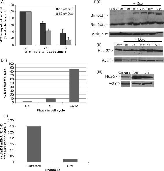
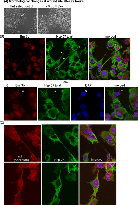
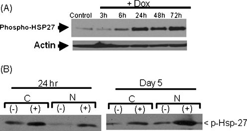
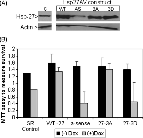
POU4F2/Brn-3b transcription factor (referred to as Brn-3b) is elevated in >60% of breast cancers and profoundly alters growth and behaviour of cancer cells by regulating distinct subsets of target genes. Previous studies showed that Brn-3b was required to maximally transactivate small heat shock protein, HSPB1/Hsp-27 (referred to as Hsp-27), and consequently, Brn-3b expression correlated well with Hsp27 levels in human breast biopsies. In these studies, we showed that Brn-3b is increased in MCF7 breast cancer cells that survive following treatment with chemotherapeutic drug doxorubicin (Dox) with concomitant increases in Hsp-27 expression. Targeting of Brn-3b using short interfering RNA reduced Hsp-27 in Dox-treated cells, suggesting that Brn-3b regulates Hsp-27 expression under these conditions. Wound healing assays showed increased Brn-3b in Dox-treated migratory cells that also express Hsp-27. Interestingly, Hsp-27 phosphorylation and cellular localisation are also significantly altered at different times following Dox treatment. Thus, phospho-Hsp-27 (p-Hsp27) protein displayed widespread distribution after 24 hrs of Dox treatment but was restricted to the nucleus after 5 days. However, in drug-resistant cells (grown in Dox for > 1 month), p-Hsp-27 was excluded from nuclei and most of the cytoplasm and appeared to be associated with the cell membrane. Studies to determine how this protein promotes survival and migration in breast cancer cells showed that the protective effects were conferred by unphosphorylated Hsp-27 protein. Thus, complex and dynamic mechanisms underlie effects of Hsp-27 protein in breast cancer cells following treatment with chemotherapeutic drugs such as Dox, and this may contribute to invasiveness and drug resistance following chemotherapy.


Figures

Fig. 1
Fig. 1
a MTT assay showing percentage of survival, following treatment of MCF7 cells with 0.5 μmol (black bar) or 1.0 μmol (grey bar) with doxorubicin for either 24 or 48 h. Loss of cell viability was expressed relative to the untreated control cells (set at 100%). b (i) Graphical representation of FACS analysis showing percentage of cells in different phases of the cell cycle following doxorubicin treatment. (ii) Expression of cyclin D1 in untreated cells (lane 1) or doxorubicin treated (Dox) cells (line 2) after 48 h. c Representative Western blot analysis showing increased (i) Brn-3b protein or (ii) HSP-27 in MCF7 cells at different times following Dox treatment. Immunoblots to detect housekeeping gene, actin, was used to assess for variability in protein loading in different sample. (iii) Immunoblot to show changes in HSP-27 protein in Dox-resistant MCF7 cells (grown in doxorubicin for up to 2 months) with actin house keeping gene controlling for protein loading. d (i) Photomicrograph showing morphological changes associated with Dox treatment of MCF7 cells with the surviving cells appearing flattened with enlarged cell body and multiple distinct processes. Immunostaining for Brn-3b (red) or HSP-27 (green) (ii) showing low protein levels in confluent, untreated cells (iii) with significant increases in Dox-treated cells. DAPI staining (blue), was used to identify cell nuclei. e Representative staining for HSP-27 protein in MCF7 transfected with (i) Brn-3b siRNA 3 (si3) that specifically silence Brn-3b. Cells expressing Brn-3b si3 that are identified by GFP (green and indicated with asterisk) show reduced HSP-27 protein expression (red). (ii) Control siRNA (NS) transfected cells stained for HSP-27 showed continued expression in GFP-positive cells to confirm specificity of si3 effects on reducing Hsp-27
Fig. 1
Fig. 1
a MTT assay showing percentage of survival, following treatment of MCF7 cells with 0.5 μmol (black bar) or 1.0 μmol (grey bar) with doxorubicin for either 24 or 48 h. Loss of cell viability was expressed relative to the untreated control cells (set at 100%). b (i) Graphical representation of FACS analysis showing percentage of cells in different phases of the cell cycle following doxorubicin treatment. (ii) Expression of cyclin D1 in untreated cells (lane 1) or doxorubicin treated (Dox) cells (line 2) after 48 h. c Representative Western blot analysis showing increased (i) Brn-3b protein or (ii) HSP-27 in MCF7 cells at different times following Dox treatment. Immunoblots to detect housekeeping gene, actin, was used to assess for variability in protein loading in different sample. (iii) Immunoblot to show changes in HSP-27 protein in Dox-resistant MCF7 cells (grown in doxorubicin for up to 2 months) with actin house keeping gene controlling for protein loading. d (i) Photomicrograph showing morphological changes associated with Dox treatment of MCF7 cells with the surviving cells appearing flattened with enlarged cell body and multiple distinct processes. Immunostaining for Brn-3b (red) or HSP-27 (green) (ii) showing low protein levels in confluent, untreated cells (iii) with significant increases in Dox-treated cells. DAPI staining (blue), was used to identify cell nuclei. e Representative staining for HSP-27 protein in MCF7 transfected with (i) Brn-3b siRNA 3 (si3) that specifically silence Brn-3b. Cells expressing Brn-3b si3 that are identified by GFP (green and indicated with asterisk) show reduced HSP-27 protein expression (red). (ii) Control siRNA (NS) transfected cells stained for HSP-27 showed continued expression in GFP-positive cells to confirm specificity of si3 effects on reducing Hsp-27
Fig. 2
Fig. 2
a Photomicrograph showing ‘wound site’ in untreated cells compared with Dox-treated cells demonstrates the distinct morphological changes following treatment (1 μmol Dox) and increased numbers of migrating cells. b Confocal images showing localisation of Brn-3b (red) or HSP-27 (green) proteins at the wound site after immunostaining. Merged picture also shows DAPI staining on the nuclei. (i) Nuclear expression of Brn-3b in cells that are positive for HSP-27 protein, which is localised primarily to the cytoplasm and in processes that make contact with other cells (indicated with asterisk). (ii) Cells without HSP-27 (marked with asterisk) do not have evident process formation compared with HSP-27-positive migrating cells. These cells are also positive Brn-3b. c Co-localisation of actin (red phalloidin staining) in processes involved in cell–cell contact, which also express HSP-27 (green). Merged picture shows nuclear (DAPI) staining also
Fig. 3
Fig. 3
a Western blot analysis showing changes in p-HSP-27 protein in MCF7 cells treated with Dox for different times (top panel). Total HSP-27 in corresponding samples are also shown (bottom panel). Invariant actin protein is used to control for differences in protein loading between the different samples shown. b Immunoblot showing p-HSP-27 in cytoplasmic (c) and nuclear ( n) fractions prepared from cells treated with Dox (+) or DMSO vehicle control (−). Cells were harvested at the times indicated after treatment for protein extraction
Fig. 4
Fig. 4
Changes in localisation of phospho-HSP-27 (red) in MCF7 cells treated with Dox for different times indicated compared with expression of the total HSP-27 (green). Representative immunostaining shows a generalised expression of total and phospho-HSP-27 after 24 h of Dox treatment. b Continued widespread distribution of total HSP-27 after 5 days Dox treatment, but phospho-HSP-27 becomes primarily nuclear after 5 days. c Restricted expression of both total and phospho-HSP-27 protein in Dox-resistant cells. d Migratory cells treated with Dox show widespread distribution of total HSP-27 but nuclear localisation of p-HSP-27
Fig. 5
Fig. 5
a Western blot analysis showing increased expression of HSP-27 following transduction of cells with AV expressing either WT, 3A or 3D mutants or reduction with antisense (AS). b Results of MTT assay to analyse changes in viability of MCF7 cells following transduction with different Hsp-27 constructs. Black bars show cell viability in untreated cells, and grey bars show changes in corresponding cells treated with Dox for 24 h. Changes in viability following Dox treatment in cells are expressed as mean (SD) of three independent experiments. Increased viability in cells with wild-type (WT) HSP-27 and non-phosphorylatable mutants are statistically significant (p< 0.05) compared with SR control. Reduction in viability in cells expressing α-sense and non-phosphorylatable mutant (27-3D) are compared to survival in absence of Dox

Similar articles

Cited by

References

    1. Arrigo AP, Suhan JP, Welch WJ. Dynamic changes in the structure and intracellular locale of the mammalian low-molecular-weight heat shock protein3. Mol Cell Biol. 1988;8:5059–5071. - PMC - PubMed
    1. Benndorf R, Hayess K, Ryazantsev S, Wieske M, Behlke J, Lutsch G. Phosphorylation and supramolecular organization of murine small heat shock protein HSP25 abolish its actin polymerization-inhibiting activity. J Biol Chem. 1994;269:20780–20784. - PubMed
    1. Bruey JM, Ducasse C, Bonniaud P, Ravagnan L, Susin SA, Diaz-latoud C, Gurbuxani S, Arrigo AP, Kroemer G, Solary E, Garrido C. Hsp27 negatively regulates cell death by interacting with cytochrome c. Nat Cell Biol. 2000;2:645–652. doi: 10.1038/35023595. - DOI - PubMed
    1. Budhram-Mahadeo V, Ndisang D, Ward T, Weber BL, Latchman DS. The Brn-3b POU family transcription factor represses expression of the BRCA-1 anti-oncogene in breast cancer cells. Oncogene. 1999;18:6684–6691. doi: 10.1038/sj.onc.1203072. - DOI - PubMed
    1. Budhram-Mahadeo V, Parker M, Latchman DS. POU transcription factors Brn-3a and Brn-3b interact with the estrogen receptor and differentially regulate transcriptional activity via an estrogen response element. Mol Cell Biol. 1998;18:1029–1041. - PMC - PubMed

Publication types

MeSH terms