Review
. 2011 Sep;109(1-3):21-32.
doi: 10.1007/s11120-011-9624-6. Epub 2011 Jan 30.

Comparative analysis of carboxysome shell proteins

Affiliations
Review

Comparative analysis of carboxysome shell proteins

James N Kinney et al. Photosynth Res. 2011 Sep.

Abstract

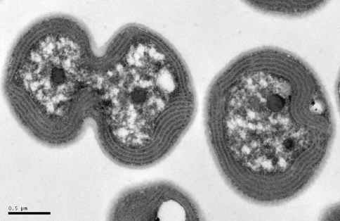
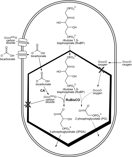
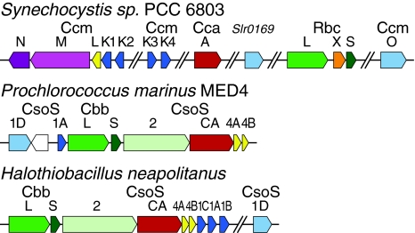
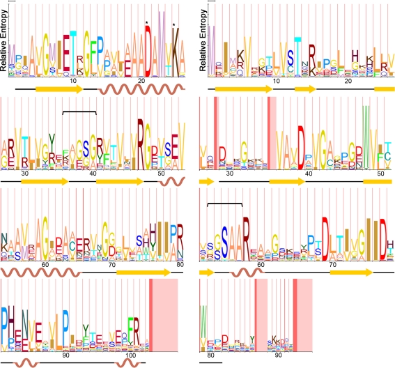
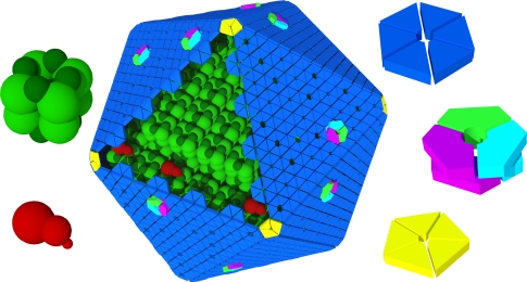
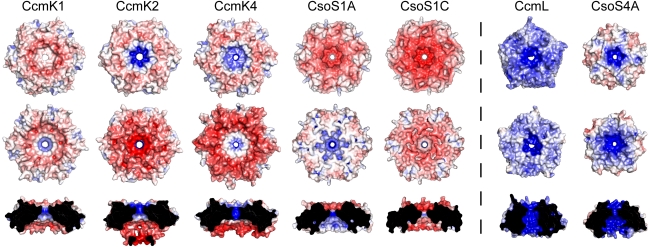
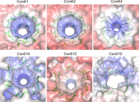
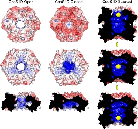
Carboxysomes are metabolic modules for CO(2) fixation that are found in all cyanobacteria and some chemoautotrophic bacteria. They comprise a semi-permeable proteinaceous shell that encapsulates ribulose-1,5-bisphosphate carboxylase/oxygenase (RuBisCO) and carbonic anhydrase. Structural studies are revealing the integral role of the shell protein paralogs to carboxysome form and function. The shell proteins are composed of two domain classes: those with the bacterial microcompartment (BMC; Pfam00936) domain, which oligomerize to form (pseudo)hexamers, and those with the CcmL/EutN (Pfam03319) domain which form pentamers in carboxysomes. These two shell protein types are proposed to be the basis for the carboxysome's icosahedral geometry. The shell proteins are also thought to allow the flux of metabolites across the shell through the presence of the small pore formed by their hexameric/pentameric symmetry axes. In this review, we describe bioinformatic and structural analyses that highlight the important primary, tertiary, and quaternary structural features of these conserved shell subunits. In the future, further understanding of these molecular building blocks may provide the basis for enhancing CO(2) fixation in other organisms or creating novel biological nanostructures.


Figures

Fig. 1
Fig. 1
Transmission electron micrograph of Synechocystis sp. PCC6803 cells showing three carboxysomes. Image courtesy of Patrick Shih, UC Berkeley
Fig. 2
Fig. 2
Schematic diagram of a cyanobacterial cell containing a carboxysome and depicting relevant metabolites that cross the cell membrane and carboxysome shell. The carboxysome-encapsulated reactions are shown. Those related to photorespiration catalyzed by RuBisCO in the presence of oxygen are shown in dashed lines
Fig. 3
Fig. 3
Three examples of carboxysome gene clusters for a β-carboxysome (top) of Synechocystis PCC 6803 and two α-carboxysomes (bottom), from the cyanobacterium Prochlorococcus marinus MED4 and from a chemoautotroph Halothiobacillus neapolitanus. Parallel diagonal lines denote large genomic segments between genes. Single-domain BMC proteins are colored dark blue; tandem-domain BMC proteins are colored light blue. Pentameric carboxysome shell proteins are colored yellow. Homologous proteins are colored similarly. Rbc and Cbb are the locus tags for RuBisCO in β- and α-carboxysomes, respectively
Fig. 4
Fig. 4
a Hidden Markov model (HMM)-logo for all unique single-domain carboxysome BMC shell proteins (CcmK1, CcmK2, CcmK3, CcmK4, CsoS1A, CsoS1B, and CsoS1C). Secondary structure of CcmK2 [Protein Data Bank (PDB) ID: 2A1B] is mapped to the corresponding positions on the logo. A horizontal bracket marks the residues lining the pore, and asterisks mark residues located at the edge of each monomer in the known structures. b HMM-logo for all Pfam03319 proteins in carboxysomes (CcmL, CsoS4A, and CsoS4B). Secondary structure of CsoS4A (PDB:2RCF) is mapped to the corresponding positions on the logo. A horizontal bracket marks the residues lining the pore. For both logos, the width of the vertical red bars is proportional to the frequency of an insertion at that position in the model. The width of the subsequent vertical pink bar is proportional to the length of that insertion [Figures prepared using MUSCLE (Edgar 2004), HMMER 3.0 (Eddy 1998), and LogoMat-M (Schuster-Bockler et al. 2004)]
Fig. 5
Fig. 5
Schematic model of the α-carboxysome assembly containing RuBisCO small (dark green) and large (green) subunits and carbonic anhydrase (red). The shell is composed of hexamers (blue), pseudohexamers (light blue, magenta, and light green), and pentamers (yellow)
Fig. 6
Fig. 6
Electrostatic comparison of structurally characterized single-domain BMC [PDB:3BN4 (CcmK1), 2A1B (CcmK2), 2A10 (CcmK4), 2G13 (CsoS1A), 3H8Y (CsoS1C)] proteins and pentameric shell proteins [PDB:2QW7 (CcmL), 2RCF (CsoS4A)]. Convex (top), concave (middle), and pore cross-section (bottom) views are shown for each structure. Red denotes negative charge; blue denotes positive charge [Figure generated with APBS Plug-in (Baker et al. 2001) for PyMOL]
Fig. 7
Fig. 7
Stereo images of superpositioned single-domain BMC monomers from the β- (blue shades) and α- (green shades) carboxysomes. The upper pair is viewed from the convex side of the protein, whereas the bottom view is rotated clockwise 90° about the x-axis from the upper view. One pore residue (Arg from CcmK4, Lys from CcmK1 and CcmK2, Phe from CsoS1A and CsoS1C) and the conserved Lys found at the edge of the hexamer are shown in yellow sticks. The regions flanked by brackets are those that display the largest structural differences between the Cso and CcmK type shell proteins
Fig. 8
Fig. 8
Conservation of all unique single-domain carboxysome BMC shell proteins mapped onto the structure of CcmK2 (PDB: 2A1B). Key residues are shown in sticks and labeled (Figure prepared using the Consurf (Ashkenazy et al. 2010) server and PyMOL)
Fig. 9
Fig. 9
Electrostatic comparison of pores from structurally characterized BMC shell proteins, viewed from the concave side. Pore residues are shown as green sticks. Red denotes negative charge; blue denotes positive charge
Fig. 10
Fig. 10
Electrostatic comparison of the two trimers of the tandem BMC-domain protein CsoS1D (PDB:3F56) and modeled representation of the “air-lock” mechanism for metabolite movement through the protein. Convex (top), concave (middle), and pore cross-section (bottom) views are shown for each of the two structures on the left. The top and bottom images of the “air-lock” mechanism are generated from the same solved stacked structure from two different orientations. The middle image is a hypothetical model generated in PyMOL by structurally aligning a copy of a closed trimer over the open trimer in the stacked structure. Red denotes negative charge and blue denotes positive charge

References

    1. Ashkenazy H, Erez E, Martz E, Pupko T, Ben-Tal N. ConSurf 2010: calculating evolutionary conservation in sequence and structure of proteins and nucleic acids. Nucleic Acids Res. 2010;38 Suppl:W529–W533. doi: 10.1093/nar/gkq399. - DOI - PMC - PubMed
    1. Badger MR, Bek EJ. Multiple rubisco forms in proteobacteria: their functional significance in relation to CO2 acquisition by the CBB cycle. J Exp Bot. 2008;59:1525–1541. doi: 10.1093/jxb/erm297. - DOI - PubMed
    1. Badger MR, Price GD. CO2 concentrating mechanisms in cyanobacteria: molecular components, their diversity and evolution. J Exp Bot. 2003;54:609–622. doi: 10.1093/jxb/erg076. - DOI - PubMed
    1. Baker SH, Lorbach SC, Rodriguez-Buey M, Williams DS, Aldrich HC, Shively JM. The correlation of the gene csoS2 of the carboxysome operon with two polypeptides of the carboxysome in Thiobacillus neapolitanus. Arch Microbiol. 1999;172:233–239. doi: 10.1007/s002030050765. - DOI - PubMed
    1. Baker NA, Sept D, Joseph S, Holst MJ, McCammon JA. Electrostatics of nanosystems: application to microtubules and the ribosome. Proc Natl Acad Sci USA. 2001;98:10037–10041. doi: 10.1073/pnas.181342398. - DOI - PMC - PubMed

Publication types

MeSH terms

LinkOut - more resources