

Crystal structure of the functional region of Uro-adherence factor A from Staphylococcus saprophyticus reveals participation of the B domain in ligand binding
- PMID: 21280131
- PMCID: PMC3048425
- DOI: 10.1002/pro.573
Crystal structure of the functional region of Uro-adherence factor A from Staphylococcus saprophyticus reveals participation of the B domain in ligand binding
Abstract
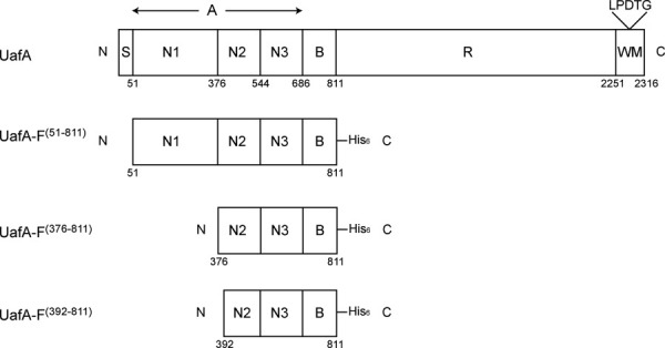
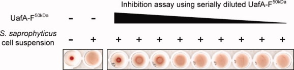
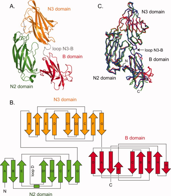
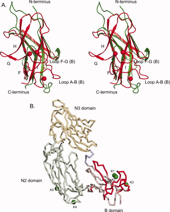
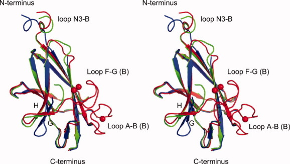
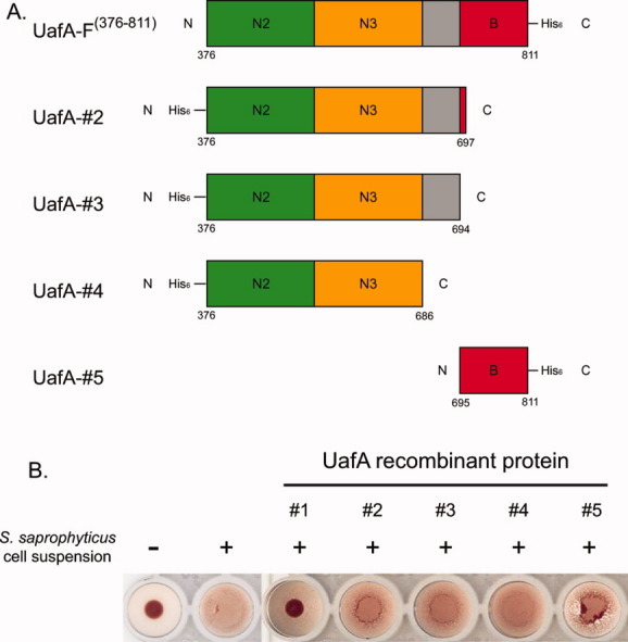
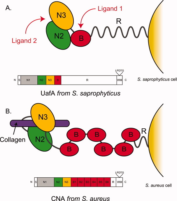
Staphylococci use cell wall-anchored proteins as adhesins to attach to host tissues. Staphylococcus saprophyticus, a uropathogenic species, has a unique cell wall-anchored protein, uro-adherence factor A (UafA), which shows erythrocyte binding activity. To investigate the mechanism of adhesion by UafA, we determined the crystal structure of the functional region of UafA at 1.5 Å resolution. The structure was composed of three domains, designated as the N2, N3, and B domains, arranged in a triangular relative configuration. Hemagglutination inhibition assay with domain-truncated mutants indicated that both N and B domains were necessary for erythrocyte binding. Based on these results, a novel manner of ligand binding in which the B domain acts as a functional domain was proposed as the adhesion mechanism of S. saprophyticus.
Copyright © 2010 The Protein Society.
Figures
Similar articles
-
UafB is a serine-rich repeat adhesin of Staphylococcus saprophyticus that mediates binding to fibronectin, fibrinogen and human uroepithelial cells.Microbiology (Reading). 2011 Apr;157(Pt 4):1161-1175. doi: 10.1099/mic.0.047639-0. Epub 2011 Jan 20. Microbiology (Reading). 2011. PMID: 21252279
-
A "dock, lock, and latch" structural model for a staphylococcal adhesin binding to fibrinogen.Cell. 2003 Oct 17;115(2):217-28. doi: 10.1016/s0092-8674(03)00809-2. Cell. 2003. PMID: 14567919
-
Aeromonas hydrophila RTX adhesin has three ligand-binding domains that give the bacterium the potential to adhere to and aggregate a wide variety of cell types.mBio. 2025 May 14;16(5):e0315824. doi: 10.1128/mbio.03158-24. Epub 2025 Apr 17. mBio. 2025. PMID: 40243363 Free PMC article.
-
Structural and functional role of Staphylococcus aureus surface components recognizing adhesive matrix molecules of the host.Future Microbiol. 2009 Dec;4(10):1337-52. doi: 10.2217/fmb.09.102. Future Microbiol. 2009. PMID: 19995192 Review.
-
A domain dictionary of trimeric autotransporter adhesins.Int J Med Microbiol. 2015 Feb;305(2):265-75. doi: 10.1016/j.ijmm.2014.12.010. Epub 2014 Dec 24. Int J Med Microbiol. 2015. PMID: 25583454 Review.
Cited by
-
Gardnerella fibrinogen-binding protein as a candidate adherence factor.Front Cell Infect Microbiol. 2025 May 8;15:1556232. doi: 10.3389/fcimb.2025.1556232. eCollection 2025. Front Cell Infect Microbiol. 2025. PMID: 40406528 Free PMC article.
-
Characterization of fibrinogen binding by glycoproteins Srr1 and Srr2 of Streptococcus agalactiae.J Biol Chem. 2013 Dec 13;288(50):35982-96. doi: 10.1074/jbc.M113.513358. Epub 2013 Oct 28. J Biol Chem. 2013. PMID: 24165132 Free PMC article.
-
Structural insights into the intermolecular interaction of the adhesin SdrC in the pathogenicity of Staphylococcus aureus.Acta Crystallogr F Struct Biol Commun. 2021 Feb 1;77(Pt 2):47-53. doi: 10.1107/S2053230X21000741. Epub 2021 Feb 2. Acta Crystallogr F Struct Biol Commun. 2021. PMID: 33620037 Free PMC article.
-
Strengthening of enterococcal biofilms by Esp.PLoS Pathog. 2022 Sep 14;18(9):e1010829. doi: 10.1371/journal.ppat.1010829. eCollection 2022 Sep. PLoS Pathog. 2022. PMID: 36103556 Free PMC article.
-
Biofilm Matrix Composition Affects the Susceptibility of Food Associated Staphylococci to Cleaning and Disinfection Agents.Front Microbiol. 2016 Jun 6;7:856. doi: 10.3389/fmicb.2016.00856. eCollection 2016. Front Microbiol. 2016. PMID: 27375578 Free PMC article.
References
-
- Wallmark G, Arremark I, Telander B. Staphylococcus saprophyticus: a frequent cause of acute urinary tract infection among female outpatients. J Infect Dis. 1978;138:791–797. - PubMed
-
- Kahlmeter G. An international survey of the antimicrobial susceptibility of pathogens from uncomplicated urinary tract infections: the ECO.SENS Project. J Antimicrob Chemother. 2003;51:69–76. - PubMed
-
- Raz R, Colodner R, Kunin CM. Who are you—Staphylococcus saprophyticus? Clin Infect Dis. 2005;40:896–898. - PubMed
MeSH terms
Substances
Associated data
- Actions
- Actions
- Actions
LinkOut - more resources
Full Text Sources
Molecular Biology Databases
