. 2011 Jan;29(1):128-40.
doi: 10.1002/stem.557.

Prospective identification and isolation of enteric nervous system progenitors using Sox2

Affiliations
Free PMC article

Prospective identification and isolation of enteric nervous system progenitors using Sox2

Tiffany A Heanue et al. Stem Cells. 2011 Jan.
Free PMC article

Abstract

The capacity to identify and isolate lineage-specific progenitor cells from developing and mature tissues would enable the development of cell replacement therapies for disease treatment. The enteric nervous system (ENS) regulates important gut functions, including controlling peristaltic muscular contractions, and consists of interconnected ganglia containing neurons and glial cells. Hirschsprung's disease (HSCR), one of the most common and best understood diseases affecting the ENS, is characterized by absence of enteric ganglia from the distal gut due to defects in gut colonization by neural crest progenitor cells and is an excellent candidate for future cell replacement therapies. Our previous microarray experiments identified the neural progenitor and stem cell marker SRY-related homoebox transcription factor 2 (Sox2) as expressed in the embryonic ENS. We now show that Sox2 is expressed in the ENS from embryonic to adult stages and constitutes a novel marker of ENS progenitor cells and their glial cell derivatives. We also show that Sox2 expression overlaps significantly with SOX10, a well-established marker of ENS progenitors and enteric glial cells. We have developed a strategy to select cells expressing Sox2, by using G418 selection on cultured gut cells derived from Sox2(βgeo/+) mouse embryos, thus allowing substantial enrichment and expansion of neomycin-resistant Sox2-expressing cells. Sox2(βgeo) cell cultures are enriched for ENS progenitors. Following transplantation into embryonic mouse gut, Sox2(βgeo) cells migrate, differentiate, and colocalize with the endogenous ENS plexus. Our studies will facilitate development of cell replacement strategies in animal models, critical to develop human cell replacement therapies for HSCR.


Figures

Figure 1
Figure 1
SRY-related homoebox transcription factor 2 (SOX2) is expressed within the mouse enteric nervous system over a broad developmental time frame. Immunohistochemistry was conducted using an anti-SOX2 antibody on cross sections through the developing (A, B) and postnatal (C, D) gut. Landmarks of the radially organized gut cross sections are noted for reference, and asterisks denote the lumen of the gut tube. (A): At E11.5, SOX2 expression is seen in the foregut endoderm (en; arrowhead) and in a punctate pattern consistent with expression in enteric neural crest-derived cells within the mesenchymal layer (me; arrows). (B): At E15.5, SOX2 expression in the midgut is observed in rings of cells corresponding to the location of the myenteric plexus (my) within the muscular layers (mu; arrows). (C): At P3, SOX2 expression in the midgut is observed in two rings, consistent with expression in the myenteric and submucosal (su) plexus layers within the muscular layers (arrows). (D): At adult stages, SOX2 expression can be seen within clusters formed between midgut muscle layers corresponding to the location of ganglia of the mature myenteric plexus (arrow). The gut epithelial layer is denoted as epi. Scale bar = 100 μm.
Figure 2
Figure 2
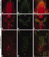
SOX2 expression is downregulated in early migratory enteric neural crest-derived cells (ENCCs), but SOX2 is expressed in ENCCs within the gut. Immunohistochemistry was conducted on sections of E9.5 (A–F) and E10.5 (G–I) embryos from the Wnt1Cre;Rosa26eYFP background, which express enhanced yellow fluorescent protein in all neural crest-derived cells (NCCs), using antibodies for SOX2 (A, D, G) and GFP (B, E, H). Merged images are shown (C, F, I). (A–C): SOX2 is expressed throughout the NT. NCCs undergoing initial migration from the neural tube downregulate SOX2 (white arrows). Faint antibody staining is detected in isolated NCCs adjacent to the ventral neural tube (yellow arrow). (D–F): At positions where ENCCs are observed colonizing the foregut (white arrowhead; foregut endoderm indicated by asterisk), SOX2 expression is not observed in ENCCs. SOX2 expression is observed within small numbers of NCCs at positions between the ventral neural tube and the dorsal aorta (da; between yellow arrows and yellow arrowhead). (G–I): At E10.5, when ENCCs are migrating extensively through the gut, SOX2 is expressed in ENCCs at all positions within the gut (yellow arrows, foregut indicated by asterisk). Coexpression of nuclear and cytoplasmic GFP (green) and nuclear SOX2 (red) is evident as green cells containing yellow/orange nuclei (see inset in [I], which corresponds to boxed region). Abbreviations: GFP, green fluorescent protein; NT, neural tube; SOX2, SRY-related homoebox transcription factor 2.
Figure 3
Figure 3
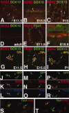
SOX2 is expressed in all SOX10-expressing enteric neural crest-derived cells and SOX2 expression is excluded from differentiated neurons. (A–F): Immunohistochemistry comparisons of SOX2 expression with expression of SOX10 (A–D) and enteric neuron markers TUJ1 (E) and HU (F) on cross sections through the E11.5 midgut (A, E), E15.5 midgut (B, F) and adult midgut (D), and peel preparations of P15 midgut outer muscle layers (C). (A–D): SOX2 and SOX10 expression is largely overlapping over a broad developmental time course. However, cells expressing only SOX2 can be identified at embryonic and early postnatal stages ([A–C], arrows). (E, F): SOX2 expression is excluded from cells expressing TUJ1 and HU. (G–T): Immunostaining on acute cultures of gut tissues from E11.5 (G, J–T), E15.5 (H), and P1 (I) wild-type animals (G–I, S, T) or Wnt1Cre;Rosa26eYFP embryos (J–R). (G–I): Comparison of SOX2 and SOX10 expression reveals that the majority of SOX2-expressing cells express SOX10 ([G–I], arrowheads; 72% at E11.5, 79% at E15.5, and 85% at P1) but that a distinct population expressing only SOX2 is evident (SOX2+ SOX10; [G–I], arrows). (J–L): Analysis of acute cultures from Wnt1Cre;Rosa26eYFP shows that all SOX2-expressing cells express green fluorescent protein (GFP) and are therefore derived from the neural crest (white arrow, white arrowhead). SOX2-expressing cells either coexpress SOX10 (white arrowhead) or do not (white arrow). GFP-expressing SOX2SOX10 cells (yellow arrowhead) have projections characteristic of differentiated neurons (yellow arrowhead). (M–O): SOX2 is expressed in neural crest-derived cells displaying punctate TUJ1 staining (arrow) but not in cells displaying a normal TUJ1 pattern ([J, K], yellow arrow). (P–R): SOX10 is expressed neither in cells displaying a punctate pattern of TUJ1 staining (white arrow) nor in cells displaying normal uniform pattern of TUJ1 staining (yellow arrow). SOX10 is expressed in cells that do not express TUJ1 (arrowhead). (S, T): Cells exhibiting a punctate pattern of TUJ1 staining also express Ki67 (arrows), and are therefore still within active phases of the cell cycle, whereas cells displaying a normal TUJ1 pattern do not express Ki67 (arrowheads), and are therefore postmitotic. Abbreviations: GFP, green fluorescent protein; SOX2, SRY-related homoebox transcription factor 2; SOX10, SRY-related homoebox transcription factor 10.
Figure 4
Figure 4
SOX2 is expressed in enteric nervous system (ENS) progenitors and glial cells. Immunostaining on acute cultures from E11.5 (A–D), E15.5 (E–H), and P1 (I–K) gut tissues using markers of ENS progenitors (SOX10, [A]), neural differentiation (HU, TUJ1, 2H3, [B–F]), and glial cell differentiation (BFABP, S100, GFAP, [G–K]). (A): SOX10 is expressed in the majority of SOX2-expressing cells at E11.5. Expression of neural markers HU, TUJ1, and 2H3 are excluded from SOX2-expressing cells at E11.5 (B–D) and E15.5 (E, F). (G, H): At E15.5, a large proportion of SOX2-expressing cells coexpress BFABP (54%, arrowheads [G]) and S100 (43%, arrowheads [H]). (I–K): At P1, almost all SOX2+ cells express markers of progressive glial cell differentiation, BFABP (99%, [I]), S100 (95%, [J]), and GFAP (32%, [K], arrowheads). Abbreviations: BFABP, brain fatty acid binding protein; GFAP, glial fibrillary acidic protein; SOX2, SRY-related homoebox transcription factor 2; SOX10, SRY-related homoebox transcription factor 10.
Figure 5
Figure 5
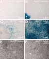
Sox2βgeo/+ mice express the βgeo transgene within the enteric nervous system (ENS), thus allowing selection of Sox2-expressing cells in culture. (A, B): β galactosidase staining on sections of E14.5 intestine (midgut; [A]) and stomach (foregut; [B]) from Sox2βgeo/+ embryos reveals expression of the βgeo transgene (blue cells) within normal sites of Sox2 expression, the ENS (arrows), and the foregut endoderm (e). (C): β galactosidase staining on primary cultures established from E14.5 Sox2βgeo/+ embryos reveals the presence of cells expressing the βgeo transgene (blue cells). (D): Primary cultures from E14.5 Sox2βgeo/+ midgut and hindgut tissue treated with G418 display a massive enrichment of cells expressing the βgeo transgene. (E, F): Bright field images of primary cultures established from E14.5 Sox2βgeo/+ reveals the presence of large flat cells that correspond to smooth muscle cells ([E], arrows), which are absent in cultures treated with G418 (F). Asterisk indicates voids present in G418-treated cultures that are likely to represent sites where G418 eliminated non-Sox2-expressing smooth muscle cells (F). LacZ indicates β galactosidase staining.
Figure 6
Figure 6
Sox2βgeo cell cultures are enriched for enteric neural crest-derived cell and glial cells and do not contain mature neurons. Immunostaining of primary cultures derived from E14.5 Sox2βgeo/+ midgut and hindgut tissues after 4 days without treatment (A, C) or treated with G418 (B, D) using markers of neural (TUJ1, [A, B]) and glial cell development (S100, [C, D]). (A): Fully differentiated TUJ1-expressing neurons (arrowhead), which are characterized by strong staining and multiple elaborated cell processes (arrows), are frequently observed in untreated cultures derived from Sox2βgeo/+ embryos. (B): Following treatment with G418, cells that express TUJ1 do so at low levels (arrowheads) and do not contain elaborated processes characteristic of differentiated neurons but contain only short processes (arrow). (C, D): The proportion of S100-expressing cells (arrowheads) is increased following treatment with G418.
Figure 7
Figure 7
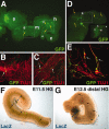
Sox2βgeo cells possess migratory and differentiation potential. (A–E): Immunostaining of cultured wild-type guts 4 days after transplantation of enhanced yellow fluorescent protein (EYFP)-expressing Sox2βgeo cells into the E11.5 gut, using GFP to detect transplanted cells and TUJ1 to detect differentiated neurons (B, C, E). (F, G): LacZ detection of Sox2βgeo cells transplanted into uncolonized regions of the E11.5 hindgut and E12.5 distal hindgut. (A): Following transplantation of a small number of cells (less than 50) into the stomach (s) of an explanted gut, EYFP-expressing cells (in green) can be observed in the midgut (m; yellow arrows in area bounded by box B) and distal hindgut (h; yellow arrows in area bounded by box C). (B, C): GFP and TUJ1 immunostaining in higher magnification view of box B and C in (A) shows transplanted GFP+ Sox2βgeo cells among endogenous TUJ1-expressing enteric neurons (in red). (D): The site of transplantation of hundreds of cells into the enteric nervous system-sparse cecum region (c), between the midgut (m) and the hindgut (h; see inset E) is identified by intense GFP expression (green arrow). Transplanted EYFP-expressing Sox2βgeo cells have migrated away from the site of transplantation (yellow arrows). (E): GFP and TUJ1 immunostaining in higher magnification view of box E in (D) shows clearly that transplanted EYFP-expressing Sox2βgeo cells express TUJ1 (arrows) and possess long processes. The transplanted TUJ1-expressing cells (in yellow) are found in close association with endogenous TUJ1-expressing neurons (in red). (F, G): Following transplantation of Sox2βgeo cells into the proximal region of the E11.5 HG (F) or E12.5 distal hindgut ([G], blue arrows, asterisks), cells migrate to the distal end of the gut segment (black arrows). Abbreviations: GFP, green fluorescent protein; HG, hindgut.

References

    1. Li L, Clevers H. Coexistence of quiescent and active adult stem cells in mammals. Science. 2010;327:542–545. - PMC - PubMed
    1. Heanue TA, Pachnis V. Enteric nervous system development and Hirschsprung's disease: Advances in genetic and stem cell studies. Nat Rev Neurosci. 2007;8:466–479. - PubMed
    1. Newgreen D, Young HM. Enteric nervous system: Development and developmental disturbances—Part 2. Pediatr Dev Pathol. 2002;5:329–349. - PubMed
    1. Newgreen D, Young HM. Enteric nervous system: Development and developmental disturbances—Part 1. Pediatr Dev Pathol. 2002;5:224–247. - PubMed
    1. Burzynski G, Shepherd IT, Enomoto H. Genetic model system studies of the development of the enteric nervous system, gut motility and Hirschsprung's disease. Neurogastroenterol Motil. 2009;21:113–127. - PMC - PubMed

Publication types

MeSH terms