. 2011 Jan 31:11:11.
doi: 10.1186/1472-6750-11-11.

Expression, secretion and surface display of a human alkaline phosphatase by the ciliate Tetrahymena thermophila

Affiliations

Expression, secretion and surface display of a human alkaline phosphatase by the ciliate Tetrahymena thermophila

Ingo Aldag et al. BMC Biotechnol. .

Abstract

Background: Tetrahymena thermophila possesses many attributes that render it an attractive host for the expression of recombinant proteins. Surface proteins from the parasites Ichthyophthirius multifiliis and Plasmodium falciparum and avian influenza virus antigen H5N1 were displayed on the cell membrane of this ciliate. Furthermore, it has been demonstrated that T. thermophila is also able to produce a functional human DNase I. The present study investigates the heterologous expression of the functional human intestinal alkaline phosphatase (hiAP) using T. thermophila and thereby presents a powerful tool for the optimization of the ciliate-based expression system.

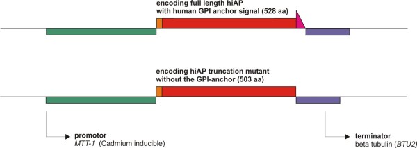
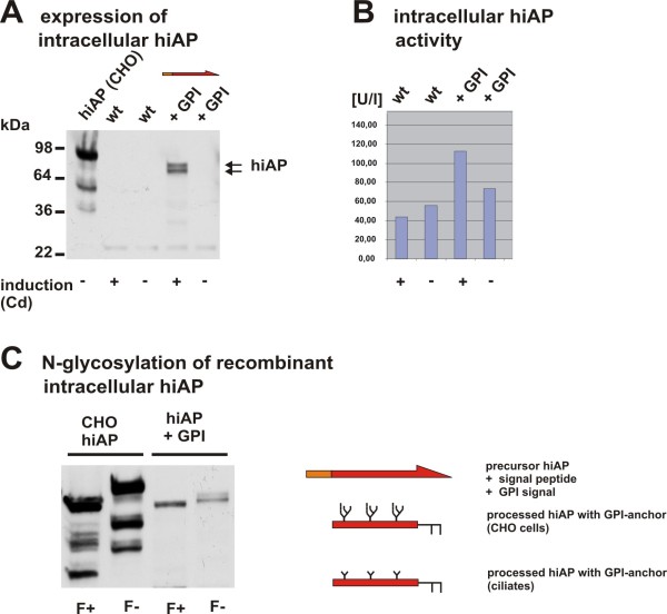
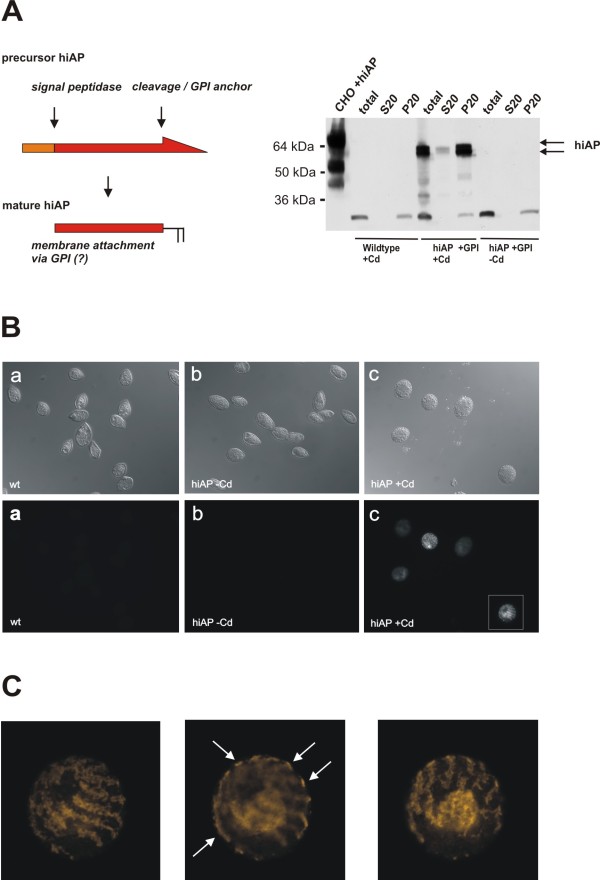
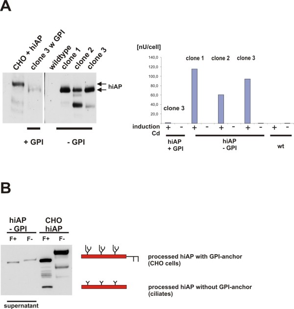
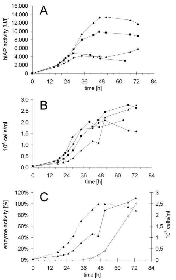
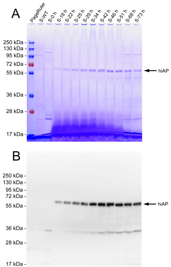
Results: Functional and full length human intestinal alkaline phosphatase was expressed by T. thermophila using a codon-adapted gene containing the native signal-peptide and GPI (Glycosylphosphatidylinositol) anchor attachment signal. HiAP activity in the cell extract of transformants suggested that the hiAP gene was successfully expressed. Furthermore, it was demonstrated that the enzyme was modified with N-glycosylation and localized on the surface membrane by the C-terminal GPI anchor. A C-terminally truncated version of hiAP lacking the GPI anchor signal peptide was secreted into the medium as an active enzyme. In a first approach to establish a high level expression system up to 14,000 U/liter were produced in a time frame of two days, which exceeds the production rate of other published expression systems for this enzyme.

Conclusions: With the expression of hiAP, not only a protein of commercial interest could be produced, but also a reporter enzyme that offers the possibility to analyze T. thermophila genes that play a role in the regulation of protein secretion. Additionally, the fact that ciliates do not secrete an endogenous alkaline phosphatase provides the possibility to use the truncated hiAP as a reporter enzyme, allowing the quantification of measures that will be necessary for further optimization of the host strains and the fermentation processes.


Figures

Figure 1
Figure 1
Structure of the used expression cassettes. Two expression cassettes were used in this study. The first expression cassette encodes the full-length hiAP precursor protein (aa 1- 528), including the N-terminal ER leader sequence (aa 1-19) and the C-terminal GPI anchor/cleavage site (aa 504-528). The second cassette encodes hiAP (aa 1- 503) without the GPI anchor/cleavage signal, consequently no GPI moiety can be added to the recombinant enzyme. The synthetic hiAP cDNA was flanked by a ~1 kb MTT1 promoter active sequence and the BTU2 terminator (~350 bp).
Figure 2
Figure 2
Characterization of recombinant full-length hiAP. A: Western blot analysis of cell extracts. In a positive clone hiAP expression was induced by the addition of cadmium to the medium. Extracts from induced cells showed a hiAP signal in the Western blot. Extracts from wild type cells (induced and non-induced) and the non-induced cells of the positive clone showed no signal. An extract from transformed CHO cells served as positive control. The double band probably corresponds to intracellular precursor hiAP with N-terminal signal peptide or to a non-cleaved GPI anchor signal. B: The data shown in the Western blot were confirmed by an alkaline phosphatase activity assay. The samples derived from wild type cells treated with and without cadmium (wt +Cd; wt -Cd) and extracts from the non-induced hiAP clone (+GPI -Cd) showed only basal activity. In contrast to this an elevated enzyme activity could be observed in cell extracts from hiAP expressing cells (+GPI +Cd), suggesting that recombinant hiAP is expressed as an active enzyme. C: We treated extracts of the hiAP expressing cells with the enzyme PNgase F (F+). Extracts from CHO cells expressing hiAP were used as a positive control. Negative controls were non-treated cell extracts (F-). The results show a significant band shift, indicating that both CHO derived as well as T. thermophila derived hiAP carries N-glycans. As expected, the band shift in ciliates is less significant due to the smaller N-glycan structure. In contrast to mammalian proteins that carry a complex N-glycosylation T. thermophila has most probably an N-glycan structure of the oligo-mannose Man3GlcNAc2 type (see scheme).
Figure 3
Figure 3
Localization of recombinant hiAP in T. thermophila cells. A: Cell fractionation experiment: Cell extracts were prepared from wild type cells and hiAP expressing cells treated with cadmium (+Cd) and without cadmium (-Cd). The whole protein preparation (total) was done without detergent and subsequently centrifuged at 20,000 × g. The insoluble fraction (pellet, P20) and the soluble fraction (supernatant, S20) were analyzed by SDS-PAGE and compared to the total fraction. Almost all of the recombinant hiAP (arrows) was found in the insoluble cell fraction, suggesting that most of the recombinant protein is proper processed and therefore membrane associated. The double band probably corresponds to intracellular precursor hiAP that is membrane attached by the signal peptide or corresponds to a non-cleaved GPI anchor signal. The scheme on the left side illustrates the processing of precursor hiAP into the mature enzyme. B: Immunofluorescence analysis: In order to confirm the cell fractionation data we performed a microscopic analysis. T. thermophila cells that express hiAP and negative controls (wild type and non-induced hiAP cells) were fixed and subsequently stained by the L-19 antiserum. The upper panel shows the Nomarski images (control) of the stained cells in the lower panel. The figure clearly illustrates that only induced cells lead to a significant staining. The cell marked with a white box was analyzed in a higher magnification. C: Detailed study of image B-c: HiAP expressing cells have a distinct staining pattern. A confocal scan through the cell demonstrated that the main hiAP signals are found on the surface of the ciliate cell (arrows). Additional signals were found in the middle of the cells. This not further characterized structure probably corresponds to transport vesicles or membranous structures that carry precursor forms of the recombinant protein. Further complete detail scans of hiAP expressing cells are shown in the two Additional files 1 and 2.
Figure 4
Figure 4
Secretion of hiAP that lacks the GPI anchor signal. A: Expression and secretion of functional recombinant hiAP. We analyzed the supernatant of three clones that were induced with cadmium. The clones carried the expression cassette that encodes hiAP without the GPI anchor signal. As control we used the clone shown in Figure 2 (clone 3 +GPI), the wild type and an extract from CHO cells that express hiAP. Clone 1, 2 and 3 show a positive signal in the Western blot analysis, the additional bands are due to protein degradation (clone 2 and 3). As expected almost no hiAP expression signal was observed in the supernatant of cells expressing hiAP +GPI (clone 3 +GPI). The enzyme activity assay (right side of the figure) confirms the data of the Western blot analysis. Clone 2 and 3 are positive, but the highest activity was found in the supernatant of clone 1. No or only few enzyme activity was found in supernatants of the wild type and in the supernatant of the clone that expresses full-length hiAP. The activity is given in nU/cell (measured activity per cell) to allow a direct comparison between the supernatant of the different clones. B: N-glycosylation of secreted hiAP. We treated the supernatant of one clone with PNGase F and analyzed the samples by SDS-PAGE and Western blot. As a control we used the extract from CHO cells that express hiAP. We found a significant band shift, suggesting that also truncated hiAP becomes glycosylated during the passage through the ER and Golgi compartments like the full-length hiAP protein. The band shift in the ciliate samples is less significant due to the smaller oligo-mannose N-glycan structure. The scheme illustrates the different post-translational modification in ciliates compared to the CHO cells.
Figure 5
Figure 5
Analysis of hiAP secretion in T. thermophila cultures. Using the Sixfors bioreactor we analyzed four 500 ml cultures that secrete hiAP without GPI in parallel. The figure shows the activity of extracellular hiAP (A) and the growing curves (B) of four independent fermentations. The cultures were induced with 10 μg/ml (circles, squares, rhombuses) or 15 μg/ml (triangles) cadmium chloride at the beginning of the experiment. Two cultures (triangles, squares) were fed with 10 times concentrated SPP medium at a rate of 2 ml/h. The maximum hiAP activity in the fed cultures (triangles, squares) was reached after 48 h whereas the maximum in the non-fed cultures was reached after 30 h. The fed culture reached about two times more hiAP activity (9,000 U/liter) compared to the unfed culture (4,500 U/liter). The induction with 15 μg/ml cadmium chloride instead of 10 μg/ml together with continuous feeding led to a further 1.5fold increase of the extracellular hiAP activity (14,000 U/liter). In a further fermentation we analyzed the secretion efficiency and compared the hiAP secretion rate to the secretion rate of the endogenous beta-glucosidase (C). The culture grew to a final titer of 2.8 × 10⁶ cells/ml (solid line, closed triangles). The beta-glucosidase activity started to increase after 36 h until the end of the fermentation (solid line, open triangles) whereas the hiAP activity only increased in the first 48 h of the cultivation reaching a stable level until the end (dashed line, closed triangles), suggesting that the secretion efficiency of the culture and the viability was not influenced by cadmium.
Figure 6
Figure 6
SDS PAGE of supernatants from different time points during fermentation. Aliquots of supernatants from different points of time (S-0 h to S-73 h) were analyzed by SDS-PAGE and subsequent Coomassie staining (A) and Western blot (B). In the Coomassie stained gel a prominent band at about 60 kDa became clearly visible which is also detectable by the L-19 antibody (B). No signals were detected before induction (S-0 h) and with a supernatant from a WT culture (S-WT).

References

    1. Collins K, Gorovsky MA. Tetrahymena thermophila. Curr Biol. 2005;15:R317–R318. doi: 10.1016/j.cub.2005.04.039. - DOI - PubMed
    1. Yao MC, Chao JL. RNA-Guided DNA Deletion in Tetrahymena: An RNAi-Based Mechanism for Programmed Genome Rearrangements. Annu Rev Genet. 2005;39:537–59. doi: 10.1146/annurev.genet.39.073003.095906. - DOI - PubMed
    1. Gorovsky MA, Yao MC, Keevert JB, Pleger GL. Isolation of micro- and macronuclei of Tetrahymena pyriformis. Methods Cell Biol. 1975;9:311–27. full_text. - PubMed
    1. Karrer KM. Tetrahymena genetics: two nuclei are better than one. Methods Cell Biol. 2000;62:127–186. full_text. - PubMed
    1. Cech TR. Beginning to understand the end of the chromosome. Cell. 2004;116:273–279. doi: 10.1016/S0092-8674(04)00038-8. - DOI - PubMed

MeSH terms

LinkOut - more resources