. 2011 Jan 31:12:31.
doi: 10.1186/1471-2474-12-31.

In-vivo generation of bone via endochondral ossification by in-vitro chondrogenic priming of adult human and rat mesenchymal stem cells

Affiliations

In-vivo generation of bone via endochondral ossification by in-vitro chondrogenic priming of adult human and rat mesenchymal stem cells

Eric Farrell et al. BMC Musculoskelet Disord. .

Abstract

Background: Bone grafts are required to repair large bone defects after tumour resection or large trauma. The availability of patients' own bone tissue that can be used for these procedures is limited. Thus far bone tissue engineering has not lead to an implant which could be used as alternative in bone replacement surgery. This is mainly due to problems of vascularisation of the implanted tissues leading to core necrosis and implant failure. Recently it was discovered that embryonic stem cells can form bone via the endochondral pathway, thereby turning in-vitro created cartilage into bone in-vivo. In this study we investigated the potential of human adult mesenchymal stem cells to form bone via the endochondral pathway.

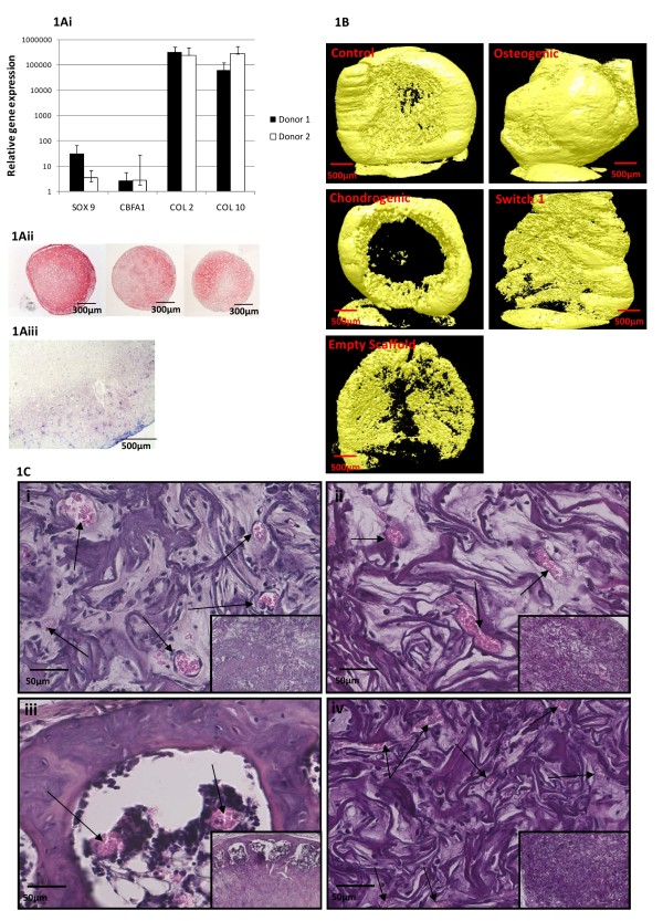
Methods: MSCs were cultured for 28 days in chondrogenic, osteogenic or control medium prior to implantation. To further optimise this process we induced mineralisation in the chondrogenic constructs before implantation by changing to osteogenic medium during the last 7 days of culture.

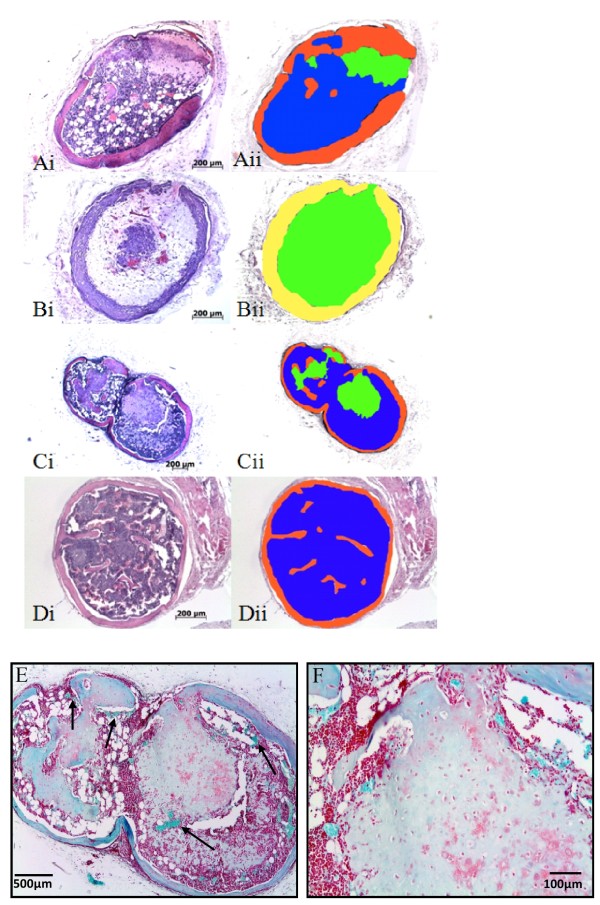
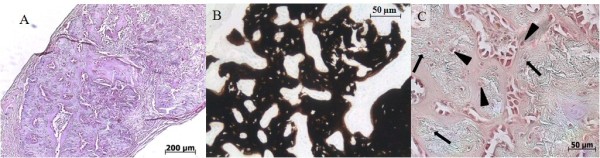
Results: After 8 weeks of subcutaneous implantation in mice, bone and bone marrow formation was observed in 8 of 9 constructs cultured in chondrogenic medium. No bone was observed in any samples cultured in osteogenic medium. Switch to osteogenic medium for 7 days prevented formation of bone in-vivo. Addition of β-glycerophosphate to chondrogenic medium during the last 7 days in culture induced mineralisation of the matrix and still enabled formation of bone and marrow in both human and rat MSC cultures. To determine whether bone was formed by the host or by the implanted tissue we used an immunocompetent transgenic rat model. Thereby we found that osteoblasts in the bone were almost entirely of host origin but the osteocytes are of both host and donor origin.

Conclusions: The preliminary data presented in this manuscript demonstrates that chondrogenic priming of MSCs leads to bone formation in vivo using both human and rat cells. Furthermore, addition of β-glycerophosphate to the chondrogenic medium did not hamper this process. Using transgenic animals we also demonstrated that both host and donor cells played a role in bone formation. In conclusion these data indicate that in-vitro chondrogenic differentiation of human MSCs could lead to an alternative and superior approach for bone tissue engineering.


Figures

Figure 1
Figure 1
Chondrogenic priming of MSCs seeded into Collagen GAG scaffolds in-vitro leads to bone formation in-vivo. Figure 1A; Chondrogenic potential was confirmed in all three donors by PCR (donors 1 and 2, expression relative to undifferentiated donor matched controls) and collagen type II immunohistochemistry (Figure 1Aii Donors 1-3). Figure 1Aiii, Toluidine Blue staining of a chondrogenically primed scaffold prior to implantation. Figure 1B Micro computed tomography of retrieved constructs (resolution 8.1 μm per pixel). The pattern of bone formation observed histologically matched closely with these images showing bone tissue at the edges of the constructs. Mineralised matrix that did not form bone was also observed in all constructs as well as empty scaffold controls. Figure 1C; Hameatoxylin and Eosin staining of bone formation in chondrogenically primed constructs (1Ciii) as compared to constructs cultured in osteogenic (Figure 1Cii) medium for 4 weeks. While osteogenically primed samples were more mineralised compared to in-vitro samples, no true bone formation was observed. Switch from chondrogenic to osteogenic medium for 7 days also prevented in-vivo bone formation (Figure 1Civ). Insets represent lower magnification images of the constructs. Arrow indicate blood vessels in each construct.
Figure 2
Figure 2
Osteogenic culture or switch prevents endochondral ossification but addition of β-glycerophosphate does not. Representative hematoxilin-eosin stained slides of implanted pellets in immune deficient mice for 8 weeks. Primed chondrogenically bone, cartilage and marrow stroma are visible (Ai). For the switch 1 condition the chondrogenic medium was replaced during the last 7 days for osteogenic medium which resulted in cartilage-like tissue in the inside and undefined tissue on the outside (Bi). For the switch 2 condition β-glycerophosphate was added during the last 7 days of culture and bone, cartilage and marrow stroma are observed (Ci). When the chondrogenic primed pellets were implanted for 14 weeks only bone and marrow stroma were visible. For quantitative analysis all pictures were pseudo colored, red (bone), blue (marrow stroma) green (cartilage), undefined tissue (yellow) (Aii, Bii, Cii, Dii). Figure 2, E and F show Safranin O staining of in vitro chondrogenically cultured pellets retrieved after 8 week in vivo. Weakly positive staining demonstrates the presence of glycosaminoglycans within a cartilage matrix being degraded to make way for bone and marrow formation which surrounds the remnants of the cartilage matrix.
Figure 3
Figure 3
Role of host and donor cells. By implanting in transgenic rats we can distinguish between donor and host with a hPLAP immunohistochemical staining. Overview of the implanted scaffold in which bone and bone marrow can be observed on hematoxilin-eosin A) and von Kossa (B) staining. All osteoblasts are stained red indicating they are from the host (C). The osteocytes however embedded in the bone are of both host (arrowheads) and donor origin (arrows).

References

    1. Meijer GJ, de Bruijn JD, Koole R, van Blitterswijk CA. Cell based bone tissue engineering in jaw defects. Biomaterials. 2008;29(21):3053–3061. doi: 10.1016/j.biomaterials.2008.03.012. - DOI - PubMed
    1. Graham SM, Leonidou A, Aslam-Pervez N, Hamza A, Panteliadis P, Heliotis M, Mantalaris A, Tsiridis E. Biological therapy of bone defects: the immunology of bone allo-transplantation. Expert Opin Biol Ther. pp. 885–901. - DOI - PubMed
    1. Akiyama M, Nakamura M. Bone regeneration and neovascularization processes in a pellet culture system for periosteal cells. Cell Transplant. 2009;18(4):443–452. doi: 10.3727/096368909788809820. - DOI - PubMed
    1. Verseijden F, Sluijs SP, Farrell E, van Neck J, Hovius S, Hofer S, van Osch G. Prevascular structures promote vascularization in engineered human adipose tissue constructs upon implantation. Cell Transplant. - PubMed
    1. Steinert AF, Ghivizzani SC, Rethwilm A, Tuan RS, Evans CH, Noth U. Major biological obstacles for persistent cell-based regeneration of articular cartilage. Arthritis Res Ther. 2007;9(3):213. doi: 10.1186/ar2195. - DOI - PMC - PubMed

Publication types

MeSH terms

Substances

LinkOut - more resources