. 2011 Jan 31:9:2.
doi: 10.1186/1477-3155-9-2.

A nanocomplex that is both tumor cell-selective and cancer gene-specific for anaplastic large cell lymphoma

Affiliations

A nanocomplex that is both tumor cell-selective and cancer gene-specific for anaplastic large cell lymphoma

Nianxi Zhao et al. J Nanobiotechnology. .

Abstract

Background: Many in vitro studies have demonstrated that silencing of cancerous genes by siRNAs is a potential therapeutic approach for blocking tumor growth. However, siRNAs are not cell type-selective, cannot specifically target tumor cells, and therefore have limited in vivo application for siRNA-mediated gene therapy.

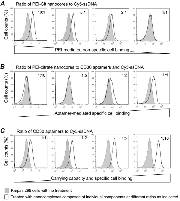
Results: In this study, we tested a functional RNA nanocomplex which exclusively targets and affects human anaplastic large cell lymphoma (ALCL) by taking advantage of the abnormal expression of CD30, a unique surface biomarker, and the anaplastic lymphoma kinase (ALK) gene in lymphoma cells. The nanocomplexes were formulated by incorporating both ALK siRNA and a RNA-based CD30 aptamer probe onto nano-sized polyethyleneimine-citrate carriers. To minimize potential cytotoxicity, the individual components of the nanocomplexes were used at sub-cytotoxic concentrations. Dynamic light scattering showed that formed nanocomplexes were ~140 nm in diameter and remained stable for more than 24 hours in culture medium. Cell binding assays revealed that CD30 aptamer probes selectively targeted nanocomplexes to ALCL cells, and confocal fluorescence microscopy confirmed intracellular delivery of the nanocomplex. Cell transfection analysis showed that nanocomplexes silenced genes in an ALCL cell type-selective fashion. Moreover, exposure of ALCL cells to nanocomplexes carrying both ALK siRNAs and CD30 RNA aptamers specifically silenced ALK gene expression, leading to growth arrest and apoptosis.

Conclusions: Taken together, our findings indicate that this functional RNA nanocomplex is both tumor cell type-selective and cancer gene-specific for ALCL cells.


Figures

Figure 1
Figure 1
Development of a tumor cell type-selective and cancer gene-specific nanocomplex for ALCL cells. A, A nano-sized carrier core structure was initially formed via aggregation of polyethyleneimine (PEI) and crosslinking with sodium citrate (PEI-citrate nanocore). The synthetic RNA-based CD30 aptamers and ALK siRNA were then incorporated onto the PEI-citrate nanocore to form the nanocomplex. B, When the functional RNA nanocomplex is added to cultures, the aptamer component will selectively target CD30-positive ALCL cells. Aptamer-mediated cell binding will facilitate intracellular delivery of the nanocomplex. The siRNA component will subsequently silence the cellular ALK gene, resulting in the growth arrest of ALCL cells.
Figure 2
Figure 2
Formulation of nanocomplex. A, Dynamic light scattering (DLS) measurement of PEI-citrate nanocores, which were formed using different 'R' ratios of citrate to PEI (charge/charge). B, Assembly of the nanocomplexes by incorporation of PEI-citrate nanocores with synthetic ALK siRNA and CD30 aptamers. The arrow indicates the addition of siRNA and aptamer components into the reaction mix. The size of the nanocomplexes formed was measured over time by DLS. C, The frequency of the nanocomplexes with different sizes was calculated. D, Nanocomplexes were incubated in cell culture medium, and the sizes were measured over time by DLS.
Figure 3
Figure 3
Cytotoxicity assays of individual nanocomplex components. A, Cultured Karpas 299 cells were treated for 48 hours with the individual components of the nanocomplex at their maximal concentrations: 100 nM CD30 aptamer, 100 nM ALK siRNA, and 4.2 μM sodium citrate, or vehicle only for the control group. Cell viability (%) was evaluated by flow cytometry using forward scatter (FSC) and side scatter (SSC) parameters as indicated. B, Karpas 299 cells were treated with PEI at concentrations of 5.48 and 1.10 μg/ml for 48 hours, and viable cells were quantified by flow cytometry, as above. C, Cell viability studies using serially diluted PEI ranging from 0.027 to 5.48 μg/ml.
Figure 4
Figure 4
Optimization of the specific cell binding and carrying capacity of the nanocomplexes. A, Synthetic Cy5-conjugated ssDNA reporter molecules were incorporated into the PEI-citrate nanocores at different ratios (moles of nitrogen in PEI to moles of phosphate in ssDNA) as indicated. Reduction of the PEI-medicated non-specific cell binding was then monitored by flow cytometry. B, To gain specific cell binding capacity, the CD30 aptamer was incorporated into PEI-citrate nanocores along with the Cy5-ssDNA reporter to form a test nanocomplex. Different ratios of PEI-citrate nanocores to total oligonucleotides (moles of nitrogen in PEI/total moles of phosphate from both the aptamer and ssDNA) were tested as indicated, while the aptamer and Cy5-ssDNA were used at a fixed ratio of 1:1 (mol/mol). The CD30 aptamer-mediated specific binding to Karpas 299 cells was confirmed using flow cytometry. C, To optimize the maximal carrying capacity, the nanocomplex was formulated using a fixed ratio of PEI-citrate nanocores to total oligonucleotides (1:1 ratio as showed in B), but the ratios of the CD30 aptamer and Cy5-ssDNA reporter were altered as indicated (mol/mol). The carrying capacity of Cy5-ssDNA reporter by the nanocomplex with specific binding to Karpas 299 cells was quantified using flow cytometry.
Figure 5
Figure 5
CD30 aptamer-mediated selective cell binding and intracellular delivery of nanocomplexes. A, Cultured Karpas 299 cells were treated with nanocomplexes containing the CD30 aptamer and Cy5-ssDNA reporter. Specific cell binding was detected by flow cytometry (top row), as well as fluorescence microscopy (bottom row) paired with light microscopy (middle row). To rule out non-specific cell binding, PEI-citrate nanocores alone or PEI-citrate nanocores containing the Cy5-ssDNA reporter (but no CD30 aptamer component) were used in control cultures. B, Cultured CD30-negative Jurkat cells were tested under the same treatment conditions. C, To assess functional biostability, the nanocomplex was pre-incubated in culture medium for up to 24 hours and changes in its cell binding capacity was kinetically monitored (%). CD30-negative Jurkat cells were used as a background binding control. D, To detect intracellular delivery, Karpas 299 cells (top row) and control Jurkat cells (bottom row) were treated with the nanocomplex containing both Cy5-ssDNA reporter and CD30 aptamer for 4 hours followed by quick nuclear staining with DAPI. As indicated, the treated cells were examined using light and confocal microscopy to visualize the DAPI-stained nuclei (blue) and the Cy5 reporter signal of the nanocomplex (red). Merged images of the DAPI-stained nuclei and Cy5 reporter signal indicate the intracellular localization of the nanocomplex.
Figure 6
Figure 6
Lymphoma cell type-dependent gene silencing by the nanocomplexes. A, Stably-expressing eGFP and luciferase Karpas 299 and Jurkat cells were used as reporters for the gene silencing studies. The cells were treated with the nanocomplexes containing eGFP siRNA along with the CD30 aptamer, non-relevant control siRNA along with CD30 aptamer, or left untreated for 2 days. Reduction of eGFP expression (%) was quantified by flow cytometry. B, Similarly, the cells were treated with nanocomplexes containing luciferase siRNA along with the CD30 aptamer for 2 days. After addition of luciferin into the cultures, the cellular luciferase activity was detected by bioluminescence scanning. C, To rule out non-specific cytotoxicity, relative viabilities (%) in the same sets of cells described in B were simultaneously examined by counting viable cell numbers. D, Cells were treated with the nanocomplexes as described in B and also in the presence of 5% or 10% fetal calf serum. After culture for 2 days at 37°C, cellular luciferase activity was detected by bioluminescence scanning.
Figure 7
Figure 7
ALK gene-silencing and growth inhibition of ALCL cells by functional RNA nanocomplexes. A, Cultured Karpas 299 cells were treated with the nanocomplex containing both ALK siRNA and CD30 aptamer for 4 days. Cellular proteins were then separated by electrophoresis and ALK fusion proteins (NPM-ALK) were detected by immunoblotting. Cellular β-actin protein expression was also measured as an internal control for gene expression. B, Cellular ALK fusion protein expression in the same set of treated Karpas 299 cells was also simultaneously detected by immunocytochemical staining. C, To study the corresponding effects on cellular proliferation and viability when the ALK gene was silenced, Karpas 299 and control Jurkat cells were treated with the nanocomplexes containing ALK siRNA or irrelevant control siRNA, or were not treated. The number of viable cells was counted under each treatment condition on days 2 and 4 post-treatment. D, To assess apoptosis, Karpas 299 cells were treated as described above for 2 days and then stained with FITC-conjugated Annexin V. The number of apoptotic cells (%) was measured by flow cytometry. ** P < 0.05.

Similar articles

Cited by

References

    1. Hannon GJ. RNA interference. Nature. 2002;418:244–251. doi: 10.1038/418244a. - DOI - PubMed
    1. Hutvagner G, Zamore PD. RNAi: nature abhors a double-strand. Curr Opin Genet Dev. 2002;12:225–232. doi: 10.1016/S0959-437X(02)00290-3. - DOI - PubMed
    1. Zamore PD. Ancient pathways programmed by small RNAs. Science. 2002;296:1265–1269. doi: 10.1126/science.1072457. - DOI - PubMed
    1. Xiang TX, Li Y, Jiang Z, Huang AL, Luo C, Zhan B, Wang PL, Tao XH. RNA interference-mediated silencing of the Hsp70 gene inhibits human gastric cancer cell growth and induces apoptosis in vitro and in vivo. Tumori. 2008;94:539–550. - PubMed
    1. Tabata T, Tsukamoto N, Fooladi AA, Yamanaka S, Furukawa T, Ishida M, Sato D, Gu Z, Nagase H, Egawa S. et al.RNA interference targeting against S100A4 suppresses cell growth and motility and induces apoptosis in human pancreatic cancer cells. Biochem Biophys Res Commun. 2009;390:475–480. doi: 10.1016/j.bbrc.2009.09.096. - DOI - PubMed

Publication types

MeSH terms

LinkOut - more resources