. 2011 Apr 1;352(1):141-51.
doi: 10.1016/j.ydbio.2011.01.027. Epub 2011 Jan 31.

Six1 regulates Grem1 expression in the metanephric mesenchyme to initiate branching morphogenesis

Affiliations

Six1 regulates Grem1 expression in the metanephric mesenchyme to initiate branching morphogenesis

Xuguang Nie et al. Dev Biol. .

Abstract

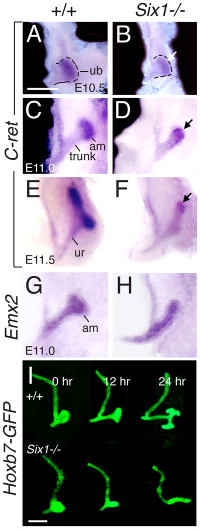
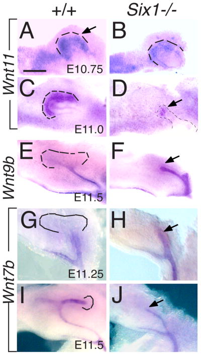
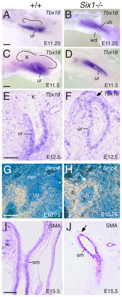
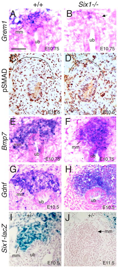
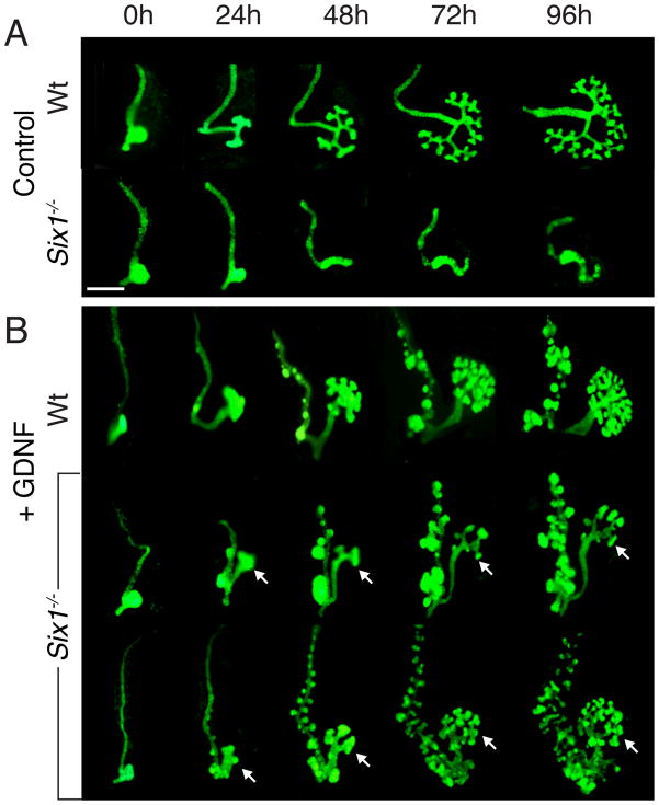
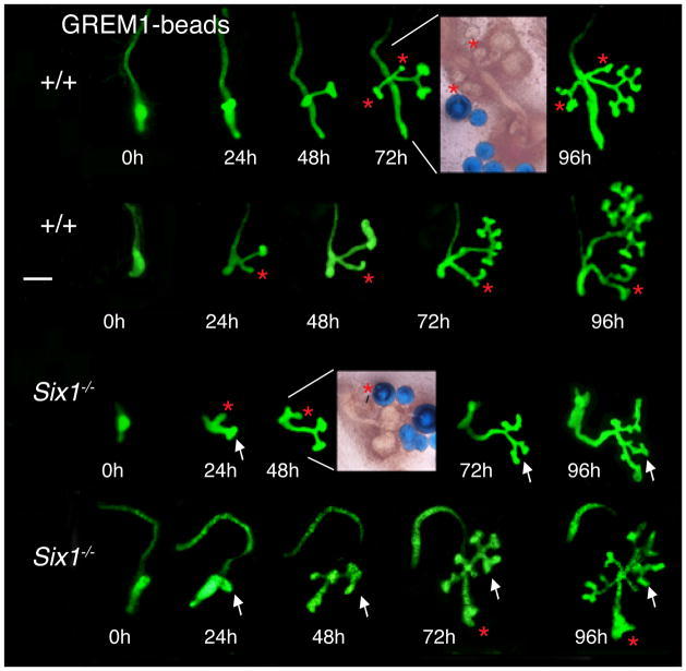
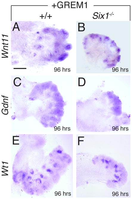
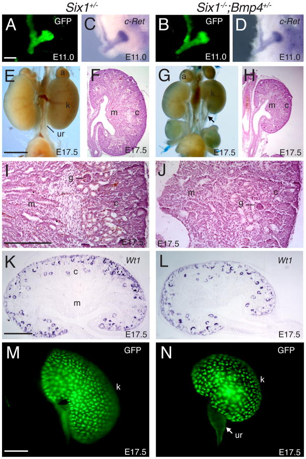
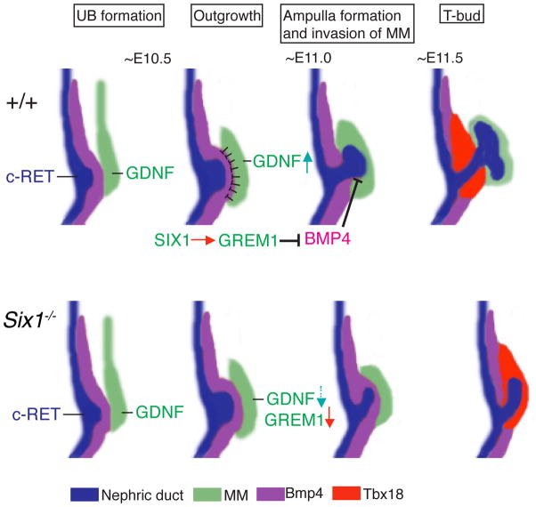
Urinary tract morphogenesis requires subdivision of the ureteric bud (UB) into the intra-renal collecting system and the extra-renal ureter, by responding to signals in its surrounding mesenchyme. BMP4 is a mesenchymal regulator promoting ureter development, while GREM1 is necessary to negatively regulate BMP4 activity to induce UB branching. However, the mechanisms that regulate the GREM1-BMP4 signaling are unknown. Previous studies have shown that Six1-deficient mice lack kidneys, but form ureters. Here, we show that the tip cells of Six1(-/-) UB fail to form an ampulla for branching. Instead, the UB elongates within Tbx18- and Bmp4-expressing mesenchyme. We find that the expression of Grem1 in the metanephric mesenchyme (MM) is Six1-dependent. Treatment of Six1(-/-) kidney rudiments with GREM1 protein restores ampulla formation and branching morphogenesis. Furthermore, we demonstrate that genetic reduction of BMP4 levels in Six1(-/-) (Six1(-/-); Bmp4(+/-)) embryos restores urinary tract morphogenesis and kidney formation. This study uncovers an essential function for Six1 in the MM as an upstream regulator of Grem1 in initiating branching morphogenesis.


Figures

Fig. 1
Fig. 1
Six1−/− UB tip fails to form ampulla. (A–H) The metanephric region was dissected out from E10.5 (A,B), E11.0 (C,D,G,H) and E11.5 (E,F) embryos and stained for c-Ret or Emx2 by whole-mount in situ hybridization. (A) Normal UB outgrowth and the formation of tip domain as labeled by c-Ret expression. (C,E) The UB tip domain swells to form an ampulla, which undergo branching to the T-bud stage at E11.5 (E), while the trunk region elongates to form the ureter. (G) E11.0 metanephric region showing Emx2 expression in ureteric stalk and ampulla. (B, D, F, H) In Six1−/− embryos, the UB outgrowth, its subdivision into the trunk and tip domains and elongation appear to occur normally, but its tip region is not dilated for the formation of ampulla. (I) Kidney rudiments expressing Hoxb7-GFP transgene were dissected from E10.5 wild-type and Six1−/− embryos and cultured in vitro for 0, 12 or 24 hour, further confirming no ampulla formation in the mutant. Abb.: am, ampulla, ub, ureteric bud, ur, ureter. Scale bars: 100 μm in A–H; 50 μm in I.
Fig. 2
Fig. 2. Altered Wnt11 expression in Six1−/− UB tips
The metanephric region was dissected out from E10.5–11.5 embryos and stained for Wnt11, Wnt9b or Wnt7b by whole-mount in situ hybridization. (A–D) Wnt11 expression in the most proximal tip domain at E10.75 (A,B) and E11.0 (C,D) in wild-type embryos and Six1−/− embryos. (E,F) Wnt9b expression in the WD and the ureteric stalk in controls and Six1−/− embryos at E11.25-11.5. (G–J) Wnt7b expression in WD and UB trunk region at E11.25 (I,J) and E11.5 (K,L) in control and Six1−/− embryos. Arrows in J and L point to the proximal end of ureteric epithelium. Dashed lines outline the UB tip regions. Scale bar: 50 μm in A–D; 80 μm in E–J.
Fig. 3
Fig. 3. Six1−/− UB elongates within Tbx18- and Bmp4-expressing mesenchyme
(A,C) Whole mount in situ hybridization of the metanephric region showing the strong Tbx18 expression domain surrounding the UB trunk region, which is ventral to the MM. Dashed lines point to UB branching within the MM. (B,D) In Six1−/− embryos, the elongating ureter is embedded within the Tbx18-expressing mesenchyme. (E,F) Section in situ at E12.5 showing Tbx18 expression surrounding the ureteral mesenchyme in control and Six1−/− embryos. Arrow points to the proximal end of the ureter in the mutant. (G,H) Radioisotope section in situ hybridization showing normal Bmp4 expression in the mesenchyme surrounding the ureteric stalk and cloaca but is absent in the metanephric mesenchyme in wild-type control (G), whereas in Six1−/− embryos, the UB tip is wrapped up by Bmp4-expressing mesenchyme (arrow). (I,J) Longitudinal sections showing SMA-positive cells surrounding the ureter in control and Six1−/− embryos at E15.5. Abb.: k, kidney, mm, metanephric mesenchyme, sm, smooth muscle, ub, ureteric bud, ur, ureter, wd, Wolffian duct. Scale bars: 50 μm in A-D; 100 μm in E–J.
Fig. 4
Fig. 4. Grem1 expression in the mesenchyme surrounding the UB tips is Six1-dependent
(A,B) Section in situ with Grem1 probe showing its expression in the mesenchyme surrounding the UB tip in normal and Six1−/− embryos. (C,D) Immunohistochemistry for pSMAD on sections from kidney region of normal and Six1−/− embryos. pSMAD-positive nuclei (brown) are present in both the mesenchyme and the epithelium of wild-type and Six1−/− embryos. pSMAD-positive cells from five sections from each MM (three kidneys) were quantified as the ratio of the pSMAD-positive cells to the total number of metanephric mesenchymal cells in the control and mutant ureters. P-values (P=0.304) were calculated using StatView t-test. Dashed lines outline the peripheral boundary of the MM. (E,F) Section in situ showing Bmp7 expression in the MM and UB in normal and Six1−/− embryos at E10.75. (G,H) Section in situ showing Gdnf expression in the MM at E10.5 normal and Six1−/− embryos. Note that the mutant MM is not as well condensed as in controls. (I) A section stained by X-gal showing Six1 expression in the MM at E10.5, (J) but its expression in the MM disappears at E11.5. Abb.: mm, metanephric mesenchyme; ub, ureteric bud. Scale bars: 50 μm.
Fig. 5
Fig. 5. Excessive GDNF is sufficient to restore not only UB branching but also tubulogenesis of the mesenchyme in Six1-deficient kidney primordia in culture
(A) Wild-type and Six1-deficient kidney primordia cultured in control medium showing normal UB outgrowth and branching and no branching in Six1−/− rudiments. (B) Control and Six1−/− kidney primordia at E10.5 were cultured in the presence of GDNF at ~200 ng/ml, many epithelial buds along the WD and overgrowth of the ureteric epithelium within 24 hours, which results in excessive branching in both wild-type and Six1-deficient kidney primordia. Note the branching and the small kidneys in GDNF-treated Six1-deficient kidney primordia. Scale bars: 100 μm.
Fig. 6
Fig. 6. Recombinant GREM1 protein is able to induce ampulla formation and branching morphogenesis in Six1−/− kidney primordia
Wild-type and Six1−/− kidney primordia expressing the Hoxb7-GFP transgene in their WD and ureteric epithelium were isolated from mouse embryos at E10.5–11.0, implanted with GREM1-soaked beads and cultured for up to 96 hours. Panels show from left to right: cultures at 0 hours, 24 hours, 48 hours, 72 hours and 96 hours (time: ±2–3 hours). White asterisks indicate ureteric buds, red asterisks indicate ectopic epithelial buds and ectopic branches. Note the branching and the small kidneys in Six1-deficient kidney primordia (white arrows). Scale bar: 100 μm.
Fig. 7
Fig. 7. Recombinant GREM1 is able to restore Wnt11 and Gdnf expression in Six1-deficient kidney rudiments in culture
Mouse kidney primordia were isolated at E10. 5–11.0, cultured for 96 hours in the presence of recombinant GREM1-soaked beads and then whole-mount stained with Wnt11 (A,B), Gdnf (C,D) and Wt1 (E,F) probes. Obvious Wnt11, Gdnf, and Wt1 expression was observed in the mutant rudiments implanted with GREM1-soaked beads and culture for 96 hours. Scale bars: 25 μm.
Fig. 8
Fig. 8. Inactivation of one copy of the Bmp4 gene in Six1−/− embryos results in formation of two small kidneys
(A,C) GFP reveals the morphology of the epithelium before fixation and (B,D) whole-mount in situ detection for the expression of the Ret receptor in control and Six1−/−;Bmp4+/− embryos at E11.25. (E,G) Gross morphology of urogenital system and (F,H) H&E staining of histological sections of kidneys of Six1+/− control (E,F) and Six1−/−;Bmp4+/− (G,H) embryos at E17.5. (I,J) Higher magnification of boxed area of F and H respectively. Developing kidney structures such as glomeruli, tubules and collecting ducts are present in the compound mutant (J). However, the patterning of the medulla (m) and cortex (c) appeared abnormal. (K,L) Kidney section of control and Six1−/−;Bmp4+/− kidney stained with Wt1 probe showing the developing nephrons. (M,N) Epithelial tracing of Hoxb7-GFP of E17.5 kidney in control and Six1−/−;Bmp4+/− animals. Arrow points to hydroureter phenotype in the compound mutant. Abb.: a, adrenal gland; c, cortex; g, glomeruli; k, kidney; m, medulla; ur, ureter. Scale bars: 50 μm in A–N; 25 μm in I,J.
Fig. 9
Fig. 9. Upregulation of Grem1 by Six1 in the MM is essential to locally restrict BMP4 signaling for the initiation of branching morphogenesis
In mice, the UB formation and outgrowth is induced by GDNF-RET signaling, which is initially expressed at normal levels in Six1−/− embryos. During this inductive period, Six1 is specifically expressed in the MM (green). Bmp4 (purple) is expressed by the mesenchyme enveloping the WD (blue) and nascent UB (blue). Grem1 transcripts are detected in the MM around the tip region of the UB, thereby locally antagonizing BMP4 in the Bmp4-expressing mesenchyme (purple) (around E10.75-11.0). This reduction of BMP4 activity by GREM1 establishes epithelial-mesenchymal interactions to enable ampulla formation of the UB and its invasion into the MM. Gdnf expression is also upregulated during this period to stimulate ampulla formation and establish WNT11-GDNF signaling for branching of the ampulla. Our results indicate that Six1 plays an essential role in lowering BMP4 activity by upregulating Grem1 expression. In Six1−/− embryos, the UB outgrowth is normal but the tip cells fail to be induced for ampulla. Instead, the UB tip is wrapped up by Tbx18- and Bmp4-expressing mesenchyme, which is the cellular source for ureteral mesenchyme (Airik et al., 2006). By responding to the signaling in the Tbx18- and Bmp4-positive mesenchyme, the tip is induced for ureter differentiation. Our results show that the balance between the levels of BMP4 activity in the Tbx18+ and Bmp4+ mesenchyme and GDNF production in the MM may also be critical for UB patterning, as excessive GDNF can restore branching as well as kidney formation in Six1−/− kidney rudiments in culture and lowering BMP4 activity in vivo can rescue branching morphogenesis and nephron formation.

Similar articles

Cited by

References

    1. Airik R, Bussen M, Singh MK, Petry M, Kispert A. Tbx18 regulates the development of the ureteral mesenchyme. J Clin Invest. 2006;116:663–74. - PMC - PubMed
    1. Airik R, Kispert A. Down the tube of obstructive nephropathies: the importance of tissue interactions during ureter development. Kidney Int. 2007;72:1459–67. - PubMed
    1. al-Awqati Q, Goldberg MR. Architectural patterns in branching morphogenesis in the kidney. Kidney Int. 1998;54:1832–42. - PubMed
    1. Blank U, Brown A, Adams DC, Karolak MJ, Oxburgh L. BMP7 promotes proliferation of nephron progenitor cells via a JNK-dependent mechanism. Development. 2009;136:3557–66. - PMC - PubMed
    1. Brenner-Anantharam A, Cebrian C, Guillaume R, Hurtado R, Sun TT, Herzlinger D. Tailbud-derived mesenchyme promotes urinary tract segmentation via BMP4 signaling. Development. 2007;134:1967–75. - PubMed

Publication types

MeSH terms