

Fast and robust extraction of hippocampus from MR images for diagnostics of Alzheimer's disease
- PMID: 21281717
- PMCID: PMC3554788
- DOI: 10.1016/j.neuroimage.2011.01.062
Fast and robust extraction of hippocampus from MR images for diagnostics of Alzheimer's disease
Abstract
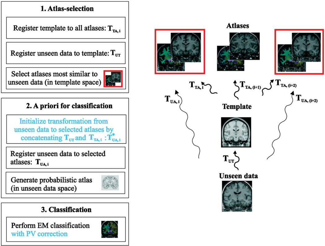
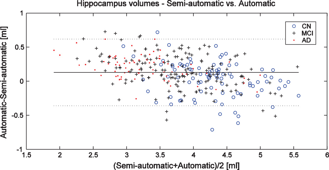
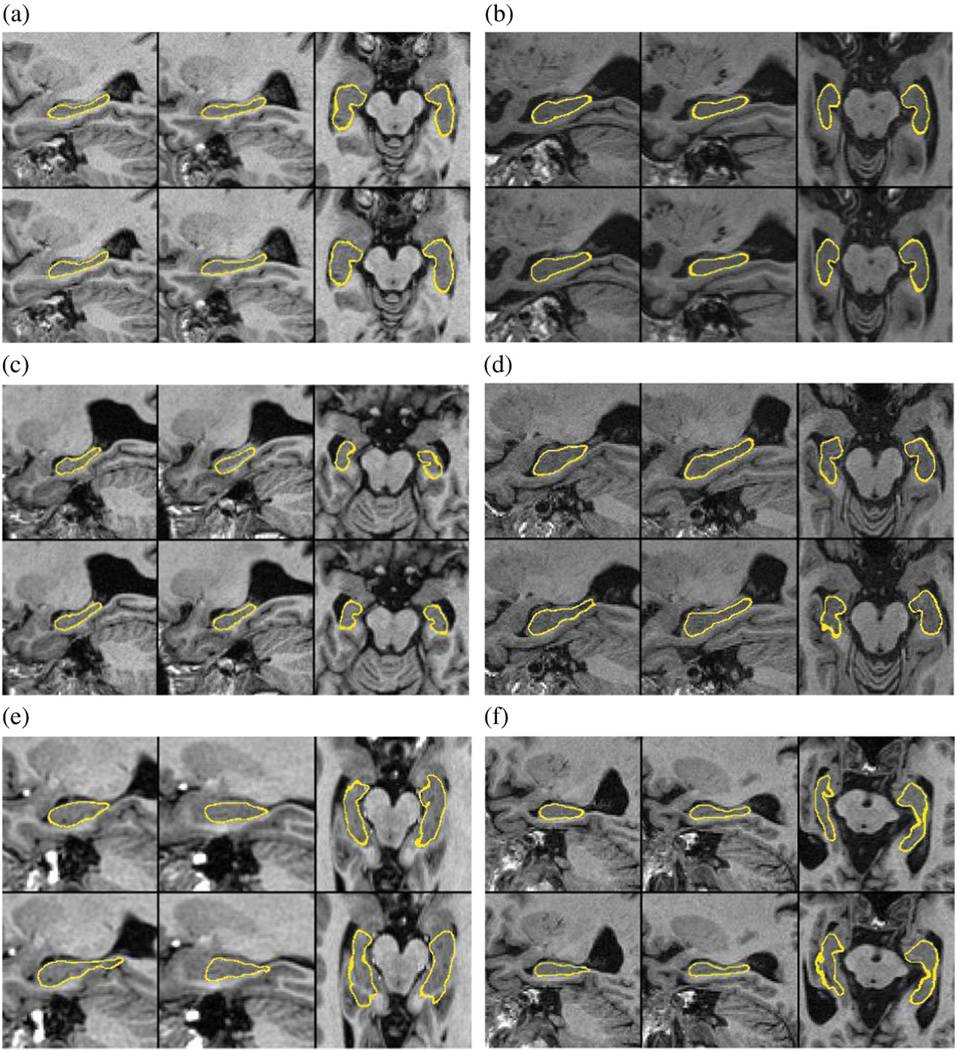
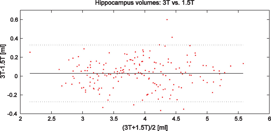
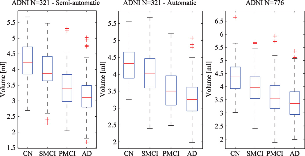
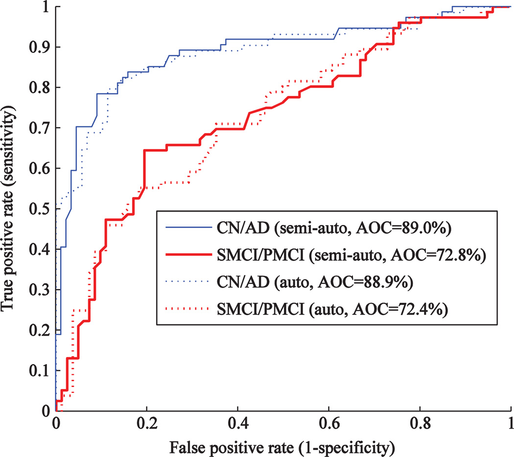
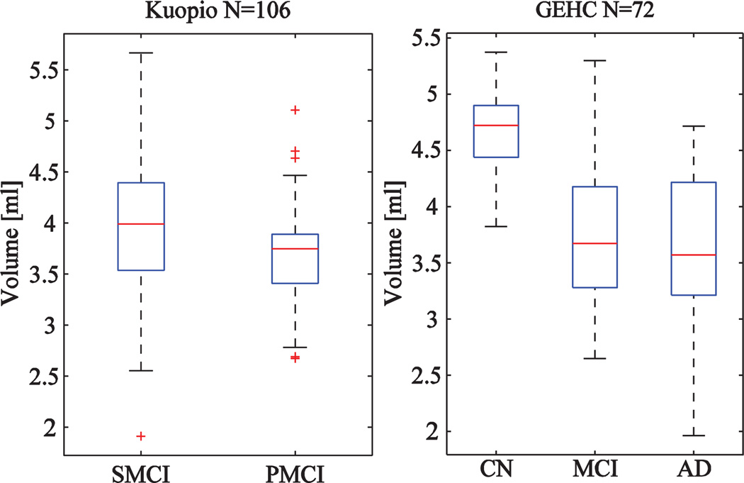
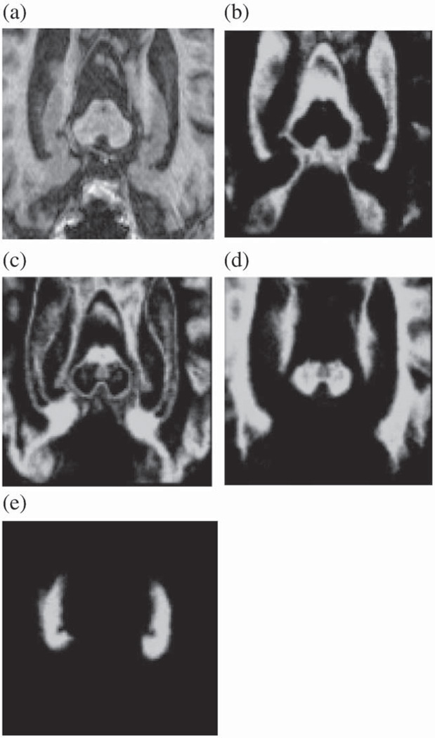
Assessment of temporal lobe atrophy from magnetic resonance images is a part of clinical guidelines for the diagnosis of prodromal Alzheimer's disease. As hippocampus is known to be among the first areas affected by the disease, fast and robust definition of hippocampus volume would be of great importance in the clinical decision making. We propose a method for computing automatically the volume of hippocampus using a modified multi-atlas segmentation framework, including an improved initialization of the framework and the correction of partial volume effect. The method produced a high similarity index, 0.87, and correlation coefficient, 0.94, with semi-automatically generated segmentations. When comparing hippocampus volumes extracted from 1.5T and 3T images, the absolute value of the difference was low: 3.2% of the volume. The correct classification rate for Alzheimer's disease and cognitively normal cases was about 80% while the accuracy 65% was obtained for classifying stable and progressive mild cognitive impairment cases. The method was evaluated in three cohorts consisting altogether about 1000 cases, the main emphasis being in the analysis of the ADNI cohort. The computation time of the method is about 2 minutes on a standard laptop computer. The results show a clear potential for applying the method in clinical practice.
Copyright © 2011 Elsevier Inc. All rights reserved.
Figures
References
-
- Babalola KO, Petenaude B, Aljabar P, Schnabel J, Kenneedy D, Crum W, Smith S, Cootes TF, Jenkinson M. Comparison and evaluation of segmentation techniques for subcortical structures in brain MRI. Med. Image Comput. Comput. Assist. Interv. MICCAI. 2008;2008(5241):409–416. - PubMed
-
- Bartko J. Measurement and reliability: statistical thinking considerations. Schizophr. Bull. 1991;17(3):483–489. - PubMed
-
- Boccardi M, Ganzola R, Duchesne S, Redolfi A, Bartzokis G, Csernansky J, deLeon MJ, Killiany RJ, Lehéricy S, Malykhin N, Pantel J, Pruessner JC, Soininen H, Jack C, Frisoni GB. Survey of segmentation protocols for hippocampal manual volumetry: preparatory phase for an EADC-ADNI harmonization protocol. Alzheimer's Demen. 2010;6:S58–S59.
-
- Cardoso MJ, Clarkson M, Ridgway G, Modat M, Fox NC, Ourselin S. Improved maximum a posteriori cortical segmentation by iterative relaxation of priors. Med. Image Comput. Comput. Assist. Interv. MICCAI. 2009;2009(5762):441–449. - PubMed
Publication types
MeSH terms
Grants and funding
LinkOut - more resources
Full Text Sources
Other Literature Sources
Medical
Research Materials
