. 2011 Feb;178(2):500-5.
doi: 10.1016/j.ajpath.2010.10.041.

The development of cutaneous neurofibromas

Affiliations

The development of cutaneous neurofibromas

Eeva-Mari Jouhilahti et al. Am J Pathol. 2011 Feb.

Abstract

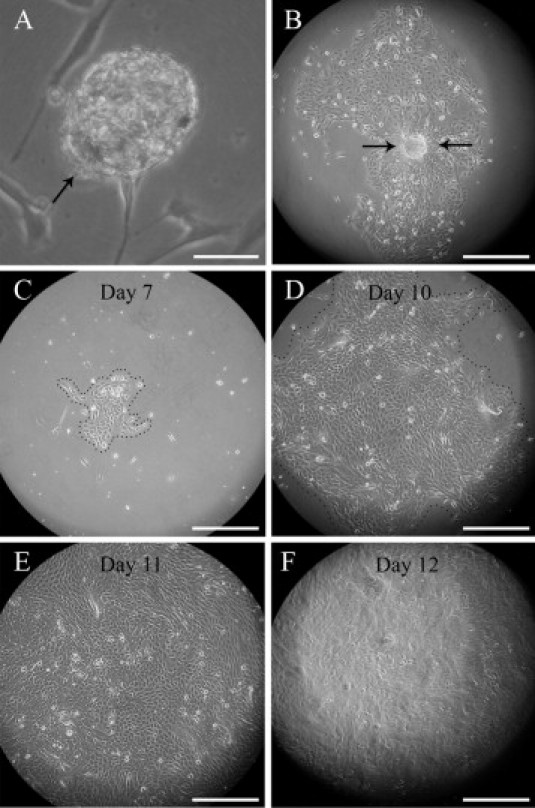
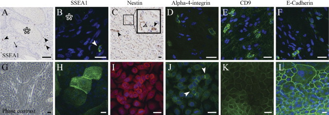
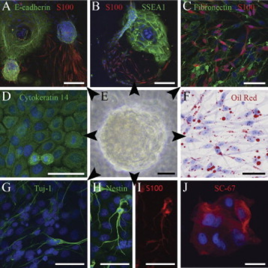
Cutaneous neurofibromas are the hallmarks of neurofibromatosis type 1 (NF1). They are composed of multiple cell types, and traditionally they are believed to arise from small nerve tributaries of the skin. A key finding in the context of this view has been that subpopulations of tumor Schwann cells harbor biallelic inactivation of the NF1 gene (NF1(-/-)). In the present study, our aim was to clarify further the pathogenesis of cutaneous neurofibromas. First, we detected cells expressing multipotency-associated biomarkers in cutaneous neurofibromas. Second, we developed a method for isolating and expanding multipotent neurofibroma-derived precursor cells (NFPs) from dissociated human cutaneous neurofibromas and used it to analyze their growth and differentiation potential. In analogy to solitary cells resident in neurofibromas, NFPs were found to express nestin and had the potential to differentiate to, at least, Schwann cells, neurons, epithelial cells, and adipocytes. Mutation analysis of the NFPs revealed that their genotype was NF1(+/-). The results led us to speculate that the development of cutaneous neurofibromas includes the recruitment of multipotent NF1(+/-) precursor cells. These cells may be derived from the multipotent cells of the hair roots, which often are intimately associated with microscopic neurofibromas.


Figures

Supplemental Figure S1
Proliferation of neurofibroma-derived cells in stem cell medium. A: A sphere of NFPs (arrow) anchored to the bottom of the culture disk. B: A single sphere has initiated the fast spreading cell sheet. CF: A 7- to 12-day time series illustrates the fast proliferation rate of progenitor cells in a growing cell sheet. At day 7 (C), the cells are still confined to a limited area, but by day 10 they fill almost the entire optical field (D), clearly exceeding it by day 11 (E). On day 12 (F), the culture displays a marked difference of appearance as it becomes multilayered. See also Supplemental Videos S1 and S2. Scale bars: 50 μm (A); 300 μm (others).
Figure 1
Figure 1
Association of hair follicles with neurofibromas at different stages. A: Cutaneous neurofibroma (diameter ∼7 mm) and numerous small, yet visible, tumor growths (left, arrows); all neurofibromas analyzed contained elements of a hair follicle (middle); three-dimensional model of a neurofibroma (right). Scale bars = 2 mm. B: Microscopic neurofibroma ensheathing a hair root (left). The rectangle depicts the approximate area further immunolabled for S100 and collagen IV; mast cells (arrowheads) are visualized by toluidine blue staining (right). Scale bars: 250 μm (left); 100 μm (S100, Collagen IV, and Tol Blue). C: Minute S100-positive neurofibroma intimately associated with hair follicle in apparently healthy looking skin of a patient with NF1. Tumor mass (arrowheads), follicular epithelium (arrows), erector pili muscle (asterisk), and mast cell (white rectangle) are shown. Scale bars = 200 μm.
Figure 2
Figure 2
Expression of SSEA1, nestin, alpha 4 integrin, CD9, and E-cadherin in neurofibroma tissue and cultured NFPs. A: Low magnification of an avidin-biotin immunolabeling shows glandular epithelium (asterisk) surrounded by neurofibroma tissue. Arrow points to the follicular epithelium within neurofibroma tissue. Arrowheads point to the SSEA1-positive cells. B: Indirect immunofluorescence labeling for SSEA1 of the same area as in A. Glandular epithelium is marked with an asterisk, and the arrowhead points to the SSEA1-positive cell. C: Small blood vessels (closed arrowheads) and a subpopulation of spindle-shaped tumor cells (open arrowhead) are positive for nestin in cutaneous neurofibroma. D: Alpha 4 integrin–positive cells shown within neurofibroma tissue. E: CD9 is expressed by a subpopulation of neurofibroma cells. F: Immunoreaction for E-cadherin. G: Phase contrast image of NFPs maintained in stem cell medium. H–L: NFPs express SSEA1, nestin, alpha 4 integrin, CD9, and E-cadherin. Arrowheads point to the positive immunoreaction for alpha 4 integrin. Collectively, the results attest to the low level of differentiation of these cells. For all fluorescence images, cell nuclei are visualized with Hoechst nuclear stain (blue). Scale bars = 100 μm (A); 20 μm (BL).
Figure 3
Figure 3
Multipotency of an NFP sphere (E) and its differentiation to mesenchymal, neural, and epithelial cells (arrowheads). A–B: Sprouting of spindle-shaped, S100-positive Schwann cells from E-cadherin–positive and SSEA1-positive NFPs. C: S100 and fibronectin labeling attest to Schwann cell and mesenchymal differentiation of NFPs. D: Cytokeratin 14 indicates epithelial differentiation. F: Differentiation to adipocytes detected with Oil Red O stain. G-I: Class III β-tubulin, nestin, and S100 indicate neuronal differentiation. J: Positive immunoreaction for neurofibromin is in line with the mutation analysis indicating the NF1+/− genotype of these cells. Schwann cells and adipocytes (C and F) were maintained in their respective differentiation mediums, whereas all other cultures were maintained in the stem cell medium. Cell nuclei are visualized with Hoechst nuclear stain. Scale bars = 200 μm (A and B); 50 μm (C–J).

References

    1. Stumpf D., Alksne J., Annegers J., Brown S., Conneally P., Housman D., Leppert M., Miller J., Moss M., Pileggi A., Rapin I., Strohman R., Swanson L., Zimmerman A. Neurofibromatosis. Conference statement. National Institutes of Health Consensus Development Conference. Arch Neurol. 1988;45:575–578. - PubMed
    1. Evans D., Komminoth P., Scheihauer B., Peltonen J. Neurofibromatosis type 1. Pathology and Genetics of Tumours of Endocrine Organs: World Health Organization Classification of Tumours. In: DeLellis R., Lioyd R., Heitz P., Eng C., editors. IARC Press; Lyon, France: 2004. pp. 243–248.
    1. Lassmann H., Jurecka W., Lassmann G., Gebhart W., Matras H., Watzek G. Different types of benign nerve sheath tumors: Light microscopy, electron microscopy and autoradiography. Virchows Arch A Pathol Anat Histopathol. 1977;375:197–210. - PubMed
    1. Peltonen J., Jaakkola S., Lebwohl M., Renvall S., Risteli L., Virtanen I., Uitto J. Cellular differentiation and expression of matrix genes in type 1 neurofibromatosis. Lab Invest. 1988;59:760–771. - PubMed
    1. Pummi K., Aho H., Laato M., Peltonen J., Peltonen S. Tight junction proteins and perineurial cells in neurofibromas. J Histochem Cytochem. 2006;54:53–61. - PubMed

Publication types