. 2011 Feb;178(2):629-39.
doi: 10.1016/j.ajpath.2010.10.030.

Attenuation of the lysosomal death pathway by lysosomal cholesterol accumulation

Affiliations

Attenuation of the lysosomal death pathway by lysosomal cholesterol accumulation

Hanna Appelqvist et al. Am J Pathol. 2011 Feb.

Abstract

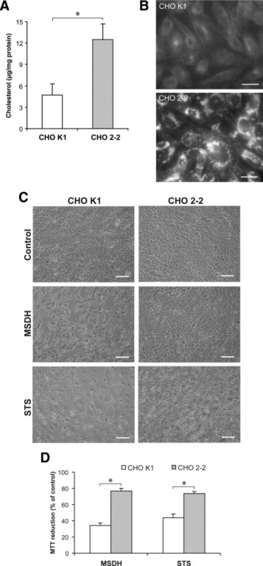
In the past decade, lysosomal membrane permeabilization (LMP) has emerged as a significant component of cell death signaling. The mechanisms by which lysosomal stability is regulated are not yet fully understood, but changes in the lysosomal membrane lipid composition have been suggested to be involved. Our aim was to investigate the importance of cholesterol in the regulation of lysosomal membrane permeability and its potential impact on apoptosis. Treatment of normal human fibroblasts with U18666A, an amphiphilic drug that inhibits cholesterol transport and causes accumulation of cholesterol in lysosomes, rescued cells from lysosome-dependent cell death induced by the lysosomotropic detergent O-methyl-serine dodecylamide hydrochloride (MSDH), staurosporine (STS), or cisplatin. LMP was decreased by pretreating cells with U18666A, and there was a linear relationship between the cholesterol content of lysosomes and their resistance to permeabilization induced by MSDH. U18666A did not induce changes in expression or localization of 70-kDa heat shock proteins (Hsp70) or antiapoptotic Bcl-2 proteins known to protect the lysosomal membrane. Induction of autophagy also was excluded as a contributor to the protective mechanism. By using Chinese hamster ovary (CHO) cells with lysosomal cholesterol overload due to a mutation in the cholesterol transporting protein Niemann-Pick type C1 (NPC1), the relationship between lysosomal cholesterol accumulation and protection from lysosome-dependent cell death was confirmed. Cholesterol accumulation in lysosomes attenuates apoptosis by increasing lysosomal membrane stability.


Figures

Figure 1
Figure 1
U18666A treatment results in cholesterol accumulation and lysosomal alterations but does not affect cell viability. Human fibroblasts were treated with 0.5 μg/ml U18666A for the indicated times. A: Intracellular cholesterol content of cells exposed to U18666A (n = 3). B: Staining of unesterified cholesterol with filipin (red) and LAMP-2 (green) in untreated cells and cells pretreated with U18666A (48 hours). The blue emission of filipin has been changed to red to better visualize the colocalization. Scale bar = 5 μm. C: Viability of cells assessed by the MTT assay (n = 4). D: Caspase-3–like activity determined using the fluorescent substrate Ac-DEVD-AMC and correlated to total protein content (n = 4). E: Flow cytometry analysis of LysoTracker Green fluorescence staining in control (green silhouette) and U18666A-treated cells (48 hours; red contour). M1 gate denotes the highly fluorescent population. F: Quantification of cells with high LysoTracker Green fluorescence (M1 gate in E) (n = 4). G: Western blot analysis of the protein expression of cathepsin D (Cat D) and LAMP-2. GAPDH was used to verify equal protein loading. One representative blot out of three is shown. H: Immunostaining (left) and quantification of BMP fluorescence (right) in fibroblasts pretreated with U18666A (48 hours) or not. Scale bar = 20 μm. Results are presented as mean values with error bars representing SD (n = 10 images). Significant difference at *P ≤ 0.05.
Figure 2
Figure 2
Fibroblasts with high cholesterol content are protected from apoptosis induced by MSDH. Human fibroblasts were pretreated with U18666A (0.5 μg/ml, 48 hours) to induce lysosomal cholesterol accumulation before exposure to the lysosomotropic detergent MSDH (15 μmol/L). A: Phase contrast images of cells exposed to MSDH for 24 hours. Scale bar = 20 μm. B: Viability of cell cultures after MSDH exposure as assessed by the MTT assay (n = 4). Viability is expressed as percentage of untreated cultures. C: Caspase-3–like activity after MSDH treatment determined using the fluorescent substrate Ac-DEVD-AMC and correlated to total protein content (n = 4). Asterisks represent statistically higher caspase-3–like activity in non-pretreated cells compared with U18666A-pretreated cells. D: Filipin staining of unesterified cholesterol in untreated cells, cells pretreated with U18666A, and cells pretreated with U18666A plus 25-HC (1 μg/ml, 48 hours). Scale bar = 20 μm. E: Viability of cultures pretreated with U18666A with or without 25-HC for 48 hours then exposed to MSDH (24 hours). Results are presented as mean values with error bars representing SD. Significant difference at *P ≤ 0.05.
Figure 3
Figure 3
Fibroblasts with high cholesterol content are less sensitive to apoptosis induced by STS and cisplatin. Human fibroblasts were pretreated with U18666A (0.5 μg/ml, 48 hours) to induce lysosomal cholesterol accumulation before exposure to STS (0.1 μmol/L, 24 hours) or cisplatin (40 μg/ml; 48 hours). A: Phase contrast images of cells exposed to STS and cisplatin. Scale bar = 20 μm. Viability and caspase-3–like activity of cell cultures after STS (B) or cisplatin (C) exposure (n = 4–8). Viability was assessed by the MTT assay and is expressed as percentage of untreated cultures. Caspase-3–like activity was determined using the fluorescent substrate Ac-DEVD-AMC and correlated to total protein content (n = 4). Results are presented as mean values with error bars representing SD. Significant difference at *P ≤ 0.05.
Figure 4
Figure 4
Augmented lysosomal cholesterol content results in increased lysosomal stability. Normal human fibroblasts were pretreated with U18666A (0.5 μg/ml, 48 hours) before exposure to MSDH (15 μmol/L). A: Immunocytochemical staining of cathepsin D and LAMP-2 after 2 hours of MSDH exposure. Scale bar = 10 μm. B: Evaluation of plasma membrane sensitivity (measured as LDH activity; n = 3) and lysosomal membrane sensitivity (measured as NAG activity; n = 3) to permeabilization with digitonin (0–200 μg/ml) in untreated (control) and U18666A-pretreated cells. Asterisk represents significantly higher NAG release in U18666A-treated cells compared with control cells. The release obtained using 200 μg/ml digitonin was regarded as maximum release (100%). C: Immunoblotting of cathepsin D (Cat D) in cytosolic fractions extracted using 20 μg/ml digitonin. LDH was used to verify equal cytosol loading. One representative blot of three is shown. D: Quantification of filipin colocalizing with LAMP-2 in human fibroblasts treated with U18666A for the indicated times (n = 10 images; each image showed at least 5 cells). Cholesterol and LAMP-2 colocalization is measured as the mean filipin fluorescence in LAMP-2–positive areas. E: Cells were pretreated with U18666A for 8, 24, and 48 hours then exposed to MSDH (1 hour). The cytosolic fraction was extracted using digitonin (20 μg/ml), and lysosomal release was measured as NAG activity. Extraction using 200 μg/ml digitonin is regarded as maximum lysosomal release. F: Correlation of lysosomal cholesterol content (as presented in D) and lysosomal stability (measured as the MSDH concentration required to result in 50% release of NAG in E). Results are presented as mean values with error bars representing SD. Significant difference at *P ≤ 0.05.
Figure 5
Figure 5
U18666A treatment is not associated with altered expression or localization of Bcl-2, Bcl-XL, Mcl-1, or Hsp70 but increased LC3-II levels. Fibroblasts were pretreated with 0.5 μg/ml U18666A for the indicated times. A: Expression of antiapoptotic Bcl-2 family proteins and Hsp70 in fibroblasts with or without U18666A treatment. GAPDH was used to verify equal protein loading. One representative blot of two is shown. B: Expression of LC3 in human fibroblasts with or without U18666A treatment. GAPDH was used to verify equal protein loading. One representative blot of three is shown. C: Immunoblotting of LC3 in human fibroblasts pretreated with 3-MA (5 mmol/L, 1 hour) followed by U18666A treatment (48 hours).
Figure 6
Figure 6
NPC-deficient CHO cells are less sensitive to MSDH- and STS-induced apoptosis than wild-type cells. A: Analysis of unesterified cholesterol content (n = 3). B: The cellular cholesterol localization using filipin in K1 (wild-type) and 2-2 (NPC-deficient) CHO cells. Scale bars = 20 μm. C: Phase-contrast images of CHO cells exposed to MSDH or STS. Scale bar = 100 μm. D: Viability of CHO cells exposed to MSDH (50 μmol/L, 24 hours) or STS (0.1 μmol/L, 24 hours). Viability is expressed as percentage of untreated cultures (n = 4). Results are presented as mean values with error bars representing SD. Significant difference at *P ≤ 0.05, as determined by nonparametric statistical testing.

References

    1. Boya P., Kroemer G. Lysosomal membrane permeabilization in cell death. Oncogene. 2008;27:6434–6451. - PubMed
    1. Firestone R.A., Pisano J.M., Bonney R.J. Lysosomotropic agents. 1. Synthesis and cytotoxic action of lysosomotropic detergents. J Med Chem. 1979;22:1130–1133. - PubMed
    1. Li W., Yuan X., Nordgren G., Dalen H., Dubowchik G.M., Firestone R.A., Brunk U.T. Induction of cell death by the lysosomotropic detergent MSDH. FEBS Lett. 2000;470:35–39. - PubMed
    1. Roberg K., Kågedal K., Öllinger K. Microinjection of cathepsin D induces caspase-dependent apoptosis in fibroblasts. Am J Pathol. 2002;161:89–96. - PMC - PubMed
    1. Johansson A.C., Appelqvist H., Nilsson C., Kågedal K., Roberg K., Öllinger K. Regulation of apoptosis-associated lysosomal membrane permeabilization. Apoptosis. 2010;15:527–540. - PMC - PubMed

Publication types

MeSH terms