. 2011 Feb;178(2):640-51.
doi: 10.1016/j.ajpath.2010.10.003.

Mast cells are an essential component of human radiation proctitis and contribute to experimental colorectal damage in mice

Affiliations

Mast cells are an essential component of human radiation proctitis and contribute to experimental colorectal damage in mice

Karl Blirando et al. Am J Pathol. 2011 Feb.

Abstract

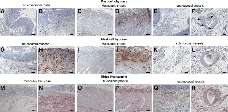
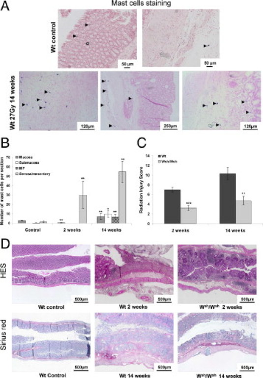
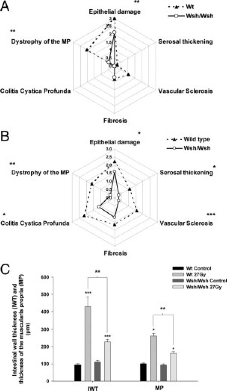
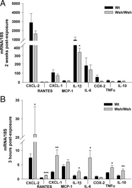
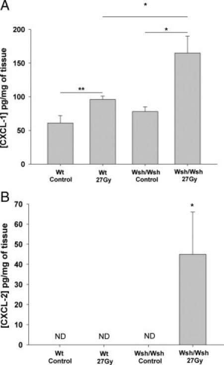
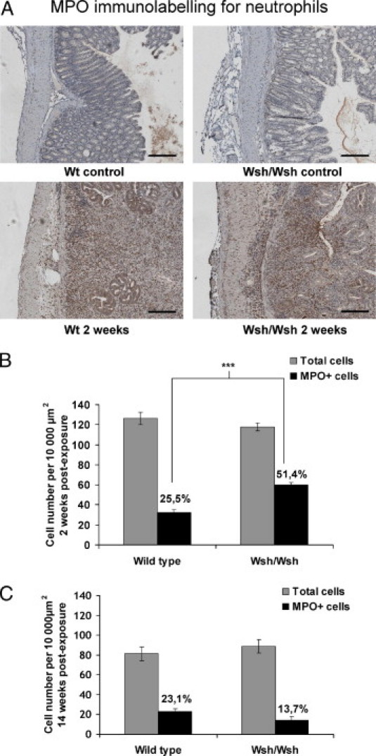
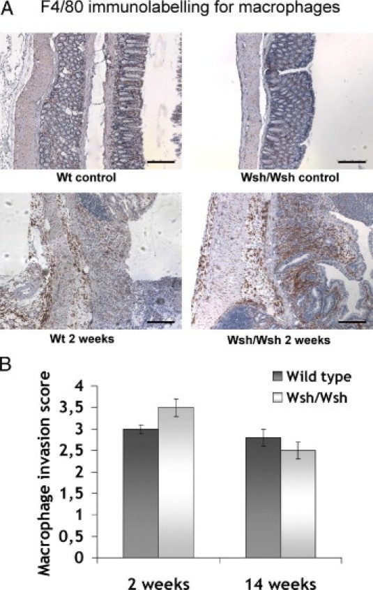
Radiation proctitis is characterized by mucosal inflammation followed by adverse chronic tissue remodeling and is associated with substantial morbidity and mortality. Mast cell hyperplasia has been associated with diseases characterized by pathological tissue remodeling and fibrosis. Rectal tissue from patients treated with radiotherapy shows mast cell hyperplasia and activation, suggesting that these cells play a role in the development of radiation-induced sequelae. To investigate the role of mast cells in radiation damage, experimental radiation proctitis was induced in a mast cell-deficient (W(sh)/W(sh)) mouse model. The colon and rectum of W(sh)/W(sh) and wild-type mice were exposed to 27-Gy single-dose irradiation and studied after 2 and 14 weeks. Irradiated rodent rectum showed mast cell hyperplasia. W(sh)/W(sh) mice developed less acute and chronic rectal radiation damage than their control littermates. Tissue protection was associated with increased tissue neutrophil influx and expression of several inflammatory mediators immediately after radiation exposure. It was further demonstrated that mast cell chymase, tryptase, and histamine could change human muscularis propria smooth muscle cells into a migrating/proliferating and proinflammatory phenotype. These data show that mast cells have deleterious effects on both acute and chronic radiation proctitis, possibly by limiting acute tissue neutrophil influx and by favoring phenotypic orientation of smooth muscle cells, thus making them active participants in the radiation-induced inflammatory process and dystrophy of the rectal wall.


Figures

Figure 1
Figure 1
Immunostaining of chymase and tryptase (red spots) in human rectal tissues. In normal tissue, chymase- and/or tryptase-positive mast cells are present in the submucosa and lamina propria (A and G). There are virtually no mast cells in the normal muscularis propria, whereas some spots are visible in the subserosa (C and I). Mast cells are absent from healthy vessel wall (E and K). Radiation injury is associated with activation and increase in mast cell numbers in the ulcerated mucosa and subjacent submucosa (B and H) and with mast cell invasion of the muscularis propria (D and J) and vascular wall [F (arrows) and L]. Sirius Red staining shows dense collagen deposition in pathological tissues in the submucosa (N), muscularis propria (P), and vessel wall (R), compared with corresponding healthy compartments (M, O, and Q, respectively). Note the absence of collagen deposition in the ulcerated mucosa (N). Scale bars = 200 μm.
Figure 2
Figure 2
A: Toluidine blue staining of mast cells in Wt tissues. Mast cells are occasionally observed in healthy mucosa and mesentery (upper panels, arrowheads). Mast cell hyperplasia is visible 14 weeks after irradiation in the MP and subserosa, as well as in the mucosa and submucosa (lower panels, arrowheads). B: Number of mast cells per tissue section in each colorectal compartment of Wt unirradiated mice (n = 5) and at 2 weeks (n = 9) and 14 weeks (n = 12) after irradiation, showing significant mast cell hyperplasia at 2 weeks in the serosa/mesentery and in all tissue compartments at 14 weeks after irradiation. *P < 0.05; **P < 0.01. C: RIS in Wt and Wsh/Wsh mice 2 and 14 weeks after exposure (n = 6–15 animals per group). **P < 0.01; ***P < 0.001. Results are presented as means ± SEM. D: Routine tissue staining with hematoxylin-eosin-saffron and collagen staining with Sirius Red. No difference in overall tissue organization was seen between Wt and Wsh/Wsh, so pictures of Wt tissues served as controls. Acute radiation injury is characterized by submucosal edema, transmural inflammation, and total loss of covering epithelium in Wt mice, whereas epithelial lining is preserved and tissue inflammation is reduced in Wsh/Wsh mice. Chronic damage shows transmural collagen deposition, mucosal disorganization, and muscular dystrophy in Wt mice, but reduced damage in Wsh/Wsh mice.
Figure 3
Figure 3
RIS parameters 2 weeks (A) and 14 weeks (B) after irradiation in Wt (dashed line; n = 9) and Wsh/Wsh mice (solid line; n = 15). *P < 0.05; **P < 0.01; ***P < 0.001. C: Intestinal wall thickness and thickness of the MP 14 weeks after injury in unirradiated mice (Wt: n = 7; Wsh/Wsh: n = 7), and in 27-Gy-irradiated Wt (n = 7) and Wsh/Wsh mice (n = 12). *P < 0.05; ***P < 0.001 compared with controls; **P < 0.01 between irradiated groups. Results are presented as means ± SEM.
Figure 4
Figure 4
mRNA expression profiles of colorectal tissues from Wt (n = 7) and Wsh/Wsh mice (n = 7) compared with respective control levels (standardized to 1) 2 weeks (A) and 3 hours (B) after irradiation. *P < 0.05; **P < 0.01; ***P < 0.001 between irradiated groups. Results are presented as means ± SEM.
Figure 5
Figure 5
Concentration of CXCL-1 (A) and CXCL-2 (B) in picograms per milligram of wet tissue in Wt and Wsh/Wsh mice 7 hours after sham (n = 6) or 27-Gy irradiation (n = 9). *P < 0.05; **P < 0.01. Results are presented as means ± SEM. ND, not detectable.
Figure 6
Figure 6
A: Immunostaining for neutrophils (in red). In unirradiated tissues, neutrophils are present in the submucosa and lamina propria in Wt, as well as in Wsh/Wsh tissues (upper panels). Irradiation induces neutrophil recruitment in all compartments in both strains (lower panels). B: Acute mucosal infiltrate in Wsh/Wsh mice (n = 8) shows a higher proportion of MPO-positive cells, compared with Wt (n = 8). ***P < 0.001. C: Late mucosal infiltrate showing no significant difference in the proportion of MPO-positive cells between Wt and Wsh/Wsh mice. Results are presented as means ± SEM.
Figure 7
Figure 7
A: Macrophages immunostaining. In unirradiated tissues, macrophages are present in the lamina propria and submucosa in Wt, as well as in Wsh/Wsh tissues (upper panels). Irradiation induces macrophage infiltration in all tissue compartments in both strains (lower panels). Scale bars = 200 μm. B: Acute and late macrophage invasion scores do not differ between strains Results are presented as means ± SEM for n = 6 (Wt) and n = 10 animals (Wsh/Wsh).
Figure 8
Figure 8
A: Activated mast cells in irradiated human rectal muscularis propria, as evidenced by chymase and tryptase immunostaining. B: Influence of chymase, tryptase, and histamine on MP-SMC proliferation. Luminescent signals corresponding to MP-SMC numbers in control conditions or 48 hours after addition of chymase, tryptase, or histamine. C: Influence of chymase, tryptase, and histamine on MP-SMC migration. Number of migrating MP-SMCs in the wound 72 hours after scratch injury, in control conditions or after addition of chymase, tryptase, or histamine. **P < 0.01; ***P < 0.001. Results are presented as means ± SEM for three independent experiments.
Figure 9
Figure 9
A: mRNA expression profile of MP-SMCs 1, 3, and 6 hours after addition of 10−5 mol/L histamine. B: IL-6 concentration (pg/ml) in MP-SMC supernatants in control conditions or 8, 16, and 24 hours after addition of 10−5 mol/L histamine. C: IL-8 concentration (pg/ml) in MP-SMC supernatants in control conditions or 8, 16, and 24 hours after addition of 10−5 mol/L histamine. *P < 0.05; **P < 0.01; ***P < 0.001. Results are presented as means ± SEM.

References

    1. Bentzen S.M., Dörr W., Anscher M.S., Denham J.W., Hauer-Jensen M., Marks L.B., Williams J. Normal tissue effects: reporting and analysis. Semin Radiat Oncol. 2003;13:189–202. - PubMed
    1. Andreyev H.J. Gastrointestinal problems after pelvic radiotherapy: the past, the present and the future. Clin Oncol (R Coll Radiol) 2007;19:790–799. - PubMed
    1. Andreyev J. Gastrointestinal symptoms after pelvic radiotherapy: a new understanding to improve management of symptomatic patients. Lancet Oncol. 2007;8:1007–1017. - PubMed
    1. Hovdenak N., Fajardo L.F., Hauer-Jensen M. Acute radiation proctitis: a sequential clinicopathologic study during pelvic radiotherapy. Int J Radiat Oncol Biol Phys. 2000;48:1111–1117. - PubMed
    1. Garg A.K., Mai W.Y., McGary J.E., Grant W.H., 3rd, Butler E.B., Teh B.S. Radiation proctopathy in the treatment of prostate cancer. Int J Radiat Oncol Biol Phys. 2006;66:1294–1305. - PubMed

Publication types

MeSH terms