. 2011 Feb;178(2):652-61.
doi: 10.1016/j.ajpath.2010.10.023.

DNA methylation suppresses expression of the urea cycle enzyme carbamoyl phosphate synthetase 1 (CPS1) in human hepatocellular carcinoma

Affiliations

DNA methylation suppresses expression of the urea cycle enzyme carbamoyl phosphate synthetase 1 (CPS1) in human hepatocellular carcinoma

Hongyan Liu et al. Am J Pathol. 2011 Feb.

Abstract

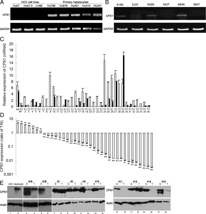
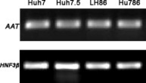
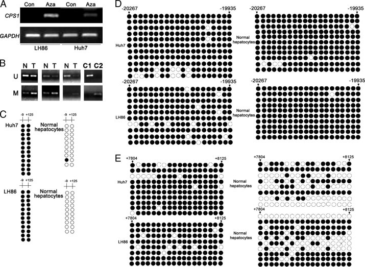
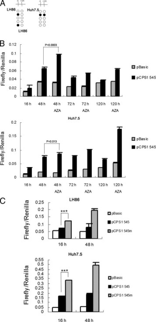
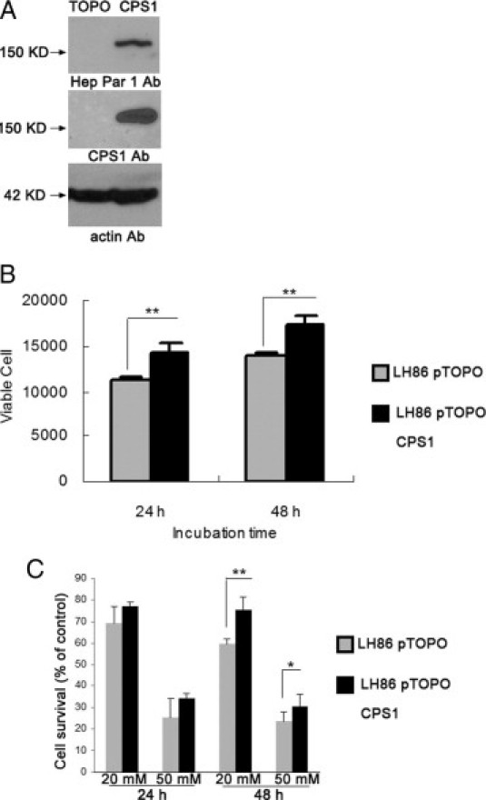
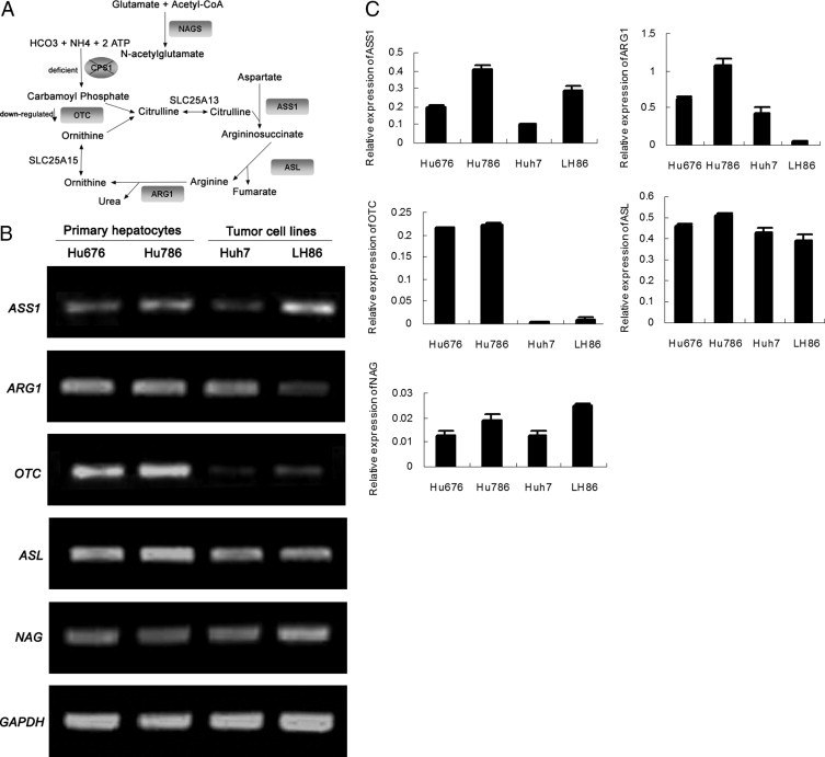
Carbamoyl phosphate synthetase 1 (CPS1) is a liver-specific, intramitochondrial, rate-limiting enzyme in the urea cycle. A previous study showed that CPS1 is the antigen for hepatocyte paraffin 1 antibody, a commonly used antibody in surgical pathology practice; and CPS1 expression appears to be down-regulated in liver cancer tissue and cell lines. The aim of this study is to understand how the CPS1 gene is regulated in liver carcinogenesis. In this report, we show that human hepatocellular carcinoma (HCC) cells do not express CPS1, whereas cultured human primary hepatocytes express abundant levels. In addition, CPS1 was silenced or down-regulated in liver tumor tissues compared with the matched noncancerous tissues. The expression of CPS1 in HCC cells was restored with a demethylation agent, 5-azacytidine. We show that two CpG dinucleotides, located near the transcription start site, and a CpG-rich region in the first intron were hypermethylated in HCC cells. The hypermethylation of the two CpG dinucleotides was also detected in HCC tumor tissues compared with noncancerous tissues. Further molecular analysis with mutagenesis indicated that the two CpG dinucleotides play a role in promoter activity of the CPS1 gene. In conclusion, our study demonstrates that DNA methylation is a key mechanism of silencing CPS1 expression in human HCC cells, and CPS1 gene hypermethylation of the two CpG dinucleotides is a potential biomarker for HCC.


Figures

Figure 1
Figure 1
Altered CPS1 expression in HCCs. A: RT-PCR analysis of CPS1 expression in HCC cell lines (ie, Huh7, Huh7.5, and LH86) and in normal primary hepatocyte cultures (ie, Hu786, Hu676, Hu401, Hu414, and Hu441). B: RT-PCR analysis of CPS1 expression in HCC tumor tissues (T) and matched nontumor liver tissues (N). The number represents deidentified case codes. Glyceraldehyde-3-phosphate dehydrogenase (GAPDH) was used as an internal control for normalization. C: Quantitative RT-PCR analysis of CPS1 transcripts in two normal liver tissues and 36 paired N and T. The number represents deidentified case codes. All data were normalized to GAPDH mRNA levels. Data are shown as the mean ± SD (n = 3). D: The ratio of CPS1 expression level between T and matched N. E: Western blot analysis of cellular extracts from cultured cells and patient tissues using Hep Par 1 antibody. Lane 1 indicates LH86 cells; 2, primary hepatocyte culture; and 3 through 22, paired N and T. Actin serves as an internal control.
Figure 2
Figure 2
The HNF3β and AAT mRNA expression levels in HCC cells. The HNF3β and AAT mRNA expression levels were analyzed by RT-PCR in HCC cell lines (ie, Huh7, Huh7.5, and LH86) and in primary hepatocyte culture (ie, Hu786).
Figure 3
Figure 3
Regulation of CPS1 expression by DNA methylation in HCCs. A: The reactivation of CPS1 gene expression by a demethylation agent in liver cancer cell lines. LH86 and Huh7 were treated with or without Aza, and CPS1 expression was examined by RT-PCR. Glyceraldehyde-3-phosphate dehydrogenase (GAPDH) was used as an internal control of the RT-PCR for normalization. B: Methylation-specific PCR analysis of CPS1 promoter in liver cancer tissues. M indicates methylated specific reaction; U, unmethylated specific reaction; C1, unmethylated control human DNA; C2, methylated control human DNA. C through E: Genomic DNA extracted from normal primary hepatocytes (ie, Hu676 and Hu786) and tumor cells (ie, Huh7 and LH86) was modified with sodium bisulfate, followed by PCR amplification, cloning, and sequencing. The DNA methylation status at each CpG site within three regions is shown. Methylated sites are indicated by filled dark circles; and unmethylated sites, empty white ones.
Figure 4
Figure 4
Functional characterization of the two CG dinucleotides in CPS1 promoter. A: LH86 and Huh7.5 cells were treated with 5 μmol/L Aza and harvested for genomic DNA extraction on 5 days of treatment. Bisulfite genomic sequencing of the CPS1 promoter [−400/145 relative to the transcription start site (+1)] was performed. The frequency of methylation at two CpG dinucleotides within the promoter is shown. B: Luciferase reporter assay of CPS1 minimal promoter activity. The LH86 and Huh7.5 cells were transfected with 600 ng of reporter gene plasmid (pGL3-CPS1 545), together with 50 ng of pRL-TK renilla luciferase plasmid (as an internal control). Twenty-four hours after transfection, the cells were treated with or without Aza for another 24, 48, or 96 hours. The cells were harvested, and luciferase activity was determined using a kit (Dual Luciferase). The data are presented as mean ± SD from three independent experiments in each cell line. C: Mutation of two CpG dinucleotides in the promoter increases CPS1 promoter activity. The LH86 and Huh7.5 cells were cotranfected with renilla luciferase vector and pGL3 CPS1-545, pGL3 CPS1-545m, or pGL3-basic control vector for 16 and 48 hours. The cells were harvested, and luciferase activity was determined using a kit (Dual Luciferase). The data are presented as mean ± SD from three independent experiments in each cell line. ***P < 0.001.
Figure 5
Figure 5
The function of CPS1 in ammonium metabolism. A: Western blot analysis of samples prepared from LH86-pTOPO control cells and from LH86-pTOPO CPS1 cells using antibodies specific against CPS1 or Hep Par 1. Actin was used as the loading control. B: The expression of CPS1 increases LH86 proliferation. C: The effects of ammonium on cell viability of LH86 cells determined by the MTS assay. LH86 was exposed to ammonium at a concentration of 20 and 50 mmol/L for 24 and 48 hours, respectively. Results are presented as the mean ± SD from three independent experiments. *P < 0.05, **P < 0.01.
Figure 6
Figure 6
Expression of urea cycle–related genes in primary hepatocytes and HCC cell lines. A: A diagram of the urea cycle. The key enzymes in the urea cycle are shown in the shadowed box. B: Expression of urea cycle–related genes was determined by RT-PCR. RNA was prepared from Hu676, Hu786, LH86, and Huh7 cells. Glyceraldehyde-3-phosphate dehydrogenase (GAPDH) was used as an internal control. C: The mRNA of the urea cycle gene was amplified by SYBR Green quantitative RT-PCR, and all values were normalized to GAPDH. CPS1, carbamoyl-phosphate; OTC, ornithine transcarbamoylase; ASS1, argininosuccinate synthetase 1; ASL, argininosuccinate lyase; ARG1, arginase 1; NAGS, N-acetylglutamate synthetase.

References

    1. Morris S.M., Jr Regulation of enzymes of urea and arginine synthesis. Annu Rev Nutr. 1992;12:81–101. - PubMed
    1. Schofield J.P. Molecular studies on an ancient gene encoding for carbamoyl-phosphate synthetase. Clin Sci (Lond) 1993;84:119–128. - PubMed
    1. Mian A., Lee B. Urea-cycle disorders as a paradigm for inborn errors of hepatocyte metabolism. Trends Mol Med. 2002;8:583–589. - PubMed
    1. Batshaw M.L. Hyperammonemia. Curr Probl Pediatr. 1984;14:1–69. - PubMed
    1. Canter J.A., Summar M.L., Smith H.B., Rice G.D., Hall L.D., Ritchie M.D., Motsinger A.A., Christian K.G., Drinkwater D.C., Jr, Scholl F.G., Dyer K.L., Kavanaugh-McHugh A.L., Barr F.E. Genetic variation in the mitochondrial enzyme carbamyl-phosphate synthetase I predisposes children to increased pulmonary artery pressure following surgical repair of congenital heart defects: a validated genetic association study. Mitochondrion. 2007;7:204–210. - PMC - PubMed

Publication types

MeSH terms