. 2011 Feb;178(2):718-23.
doi: 10.1016/j.ajpath.2010.10.016.

IgG autoantibodies against desmocollin 3 in pemphigus sera induce loss of keratinocyte adhesion

Affiliations

IgG autoantibodies against desmocollin 3 in pemphigus sera induce loss of keratinocyte adhesion

David Rafei et al. Am J Pathol. 2011 Feb.

Erratum in

  • Am J Pathol. 2013 Feb;182(2):610

Abstract

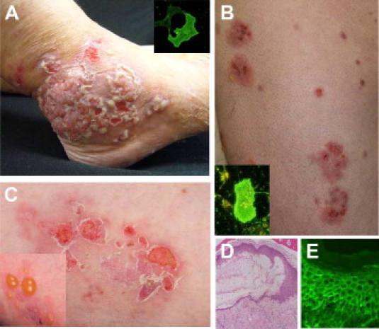
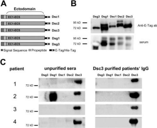
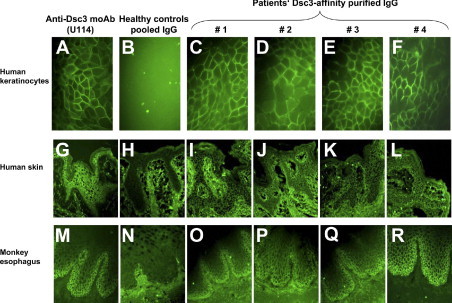
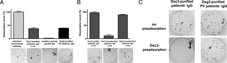
Pemphigus is considered an autoimmune bullous skin disorder associated with IgG against the desmosomal components, desmoglein 3 (Dsg3) and desmoglein 1 (Dsg1). This concept is supported by the in vitro and in vivo pathogenicity of anti-Dsg3/Dsg1 IgG and the mucosal blistering phenotype of mice with a genetic deficiency of Dsg3. Mice deficient for another desmosomal adhesion molecule, desmocollin 3 (Dsc3), show a similar pemphigus phenotype, and we investigated the pathogenicity of Dsc3-reactive IgG autoantibodies that were identified previously in a subset of patients with atypical pemphigus. We here demonstrate that IgG against Dsc3 causes loss of adhesion of epidermal keratinocytes. Specifically, IgG against Dsc3 was purified from Dsc3-reactive pemphigus sera by affinity column chromatography using recombinant human Dsc3. Affinity purified IgG was functionally active and did not only react with recombinant Dsc3 but also with epidermis and cultured human keratinocytes. Moreover, Dsc3-reactive IgG induced loss of adhesion of epidermal keratinocytes in a dispase-based keratinocyte dissociation assay that was reversed on pre-adsorption with human Dsc3 but not Dsg3. These findings demonstrate that IgG autoantibodies against an additional component of the desmosomes, Dsc3, induce loss of keratinocyte adhesion and thus may contribute to blister formation in pemphigus.


Figures

Figure 1
Figure 1
Clinical phenotype of representative patients with atypical pemphigus. Clinical pictures of two representative patients with atypical pemphigus (pemphigus vegetans and pemphigus herpetiformis), illustrating hypertrophic verrucous plaques with pustules and erosions of the foot (A) and multiple erythematous herpetiform erosions of the trunk and extremities (B, C). Insets of A and B show IgG reactivity of the patients' sera with human desmocollin 3-transfected COS-7 cells. Histopathology of lesional skin reveals intraepidermal loss of keratinocyte adhesion at the suprabasilar level (D). Direct immunofluorescence shows IgG deposits on the surface of epidermal keratinocytes (E).
Figure 2
Figure 2
Affinity purification of desmocollin 3 (Dsc3)-reactive IgG autoantibodies from four patients with atypical pemphigus. A: Scheme of the recombinant proteins applied in this study, ie, the entire ectodomains of Dsc1, Dsc2, Dsc3, desmoglein 1 (Dsg1), and desmoglein 3 (Dsg3) linked to E- and 6xHis-Tag. B: Detection of the recombinant proteins by immunoblot analysis using an anti-E-tag monoclonal antibody (upper panel) or serum of a patient with paraneoplastic pemphigus (lower panel). C: Immunoblot analysis revealing IgG reactivity of the four atypical pemphigus sera, with the recombinant proteins before (left panels) and after (right panels) affinity purification, using Dsc3 columns. Patient 2 shows additional IgG reactivity with Dsg1.
Figure 3
Figure 3
Reactivity of the purified desmocollin 3 (Dsc3)-reactive IgG autoantibodies with cultured human keratinocytes, human epidermis, and monkey esophagus. Dsc3 affinity-purified IgG from four patients, with atypical pemphigus, reveal intercellular reactivity with cultured human keratinocytes (HaCaT cells) [patients 1–4 (C–F)], human epidermis (I–L) and monkey esophagus (O–R). Monoclonal anti-Dsc3 antibody (clone U114) (A, G, and M) and pooled IgG of healthy control sera (B, H, and N) served as positive and negative controls, respectively.
Figure 4
Figure 4
Desmocollin 3 (Dsc3)-affinity purified IgG from four patients with atypical pemphigus induces loss of keratinocyte adhesion. Results of the dispase-based keratinocyte dissociation assay using primary human epidermal keratinocytes are illustrated. Keratinocyte monolayers were incubated with the monoclonal anti-Dsc3 IgG antibody (clone U114), Dsc3-affinity purified IgG from the four atypical pemphigus patients, pooled IgG from healthy donors, or desmoglein 3 (Dsg3)-affinity purified IgG from a pemphigus vulgaris patient. A: Quantification of the keratinocyte fragments is depicted as a relative dissociation score. The maximal number of keratinocyte fragments obtained after incubation with the monoclonal anti-Dsc3 IgG antibody (clone U114) is set as 100%. Representative images out of three independent experiments are shown below the diagram. B: Number of keratinocyte fragments after incubation with Dsc3-affinity purified IgG from four atypical pemphigus patients is illustrated as a dissociation score of 100% (B, left column). The induction of keratinocyte dissociation is Dsc3-specific because pre-adsorption of the Dsc3-affinity purified IgG fraction with recombinant Dsc3 (B, middle column and C, left panel) but not with human Dsg3 (B, right column) blocks keratinocyte dissociation. To exclude non-specific binding of IgG to recombinant Dsc3 protein, Dsg3-purified IgG from a pemphigus vulgaris patient was pre-adsorbed with Dsc3 protein (C, right panels). Dsg3 affinity purified IgG induces loss of keratinocyte adhesion (A and C) that is not inhibited by pre-adsorption with human Dsc3 protein (C, right panels).

Similar articles

Cited by

References

    1. Amagai M., Klaus-Kovtun V., Stanley J.R. Autoantibodies against a novel epithelial cadherin in pemphigus vulgaris, a disease of cell adhesion. Cell. 1991;67:869–877. - PubMed
    1. Hertl M., Eming R., Veldman C. T cell control in autoimmune bullous skin disorders. J Clin Invest. 2006;116:1159–1166. - PMC - PubMed
    1. Amagai M., Hashimoto T., Shimizu N., Nishikawa T. Absorption of pathogenic autoantibodies by the extracellular domain of pemphigus vulgaris antigen (Dsg3) produced by baculovirus. J Clin Invest. 1994;94:59–67. - PMC - PubMed
    1. Mahoney M.G., Wang Z., Rothenberger K., Koch P.J., Amagai M., Stanley J.R. Explanations for the clinical and microscopic localization of lesions in pemphigus foliaceus and vulgaris. J Clin Invest. 1999;103:461–468. - PMC - PubMed
    1. Amagai M., Koch P.J., Nishikawa T., Stanley J.R. Pemphigus vulgaris antigen (desmoglein 3) is localized in the lower epidermis, the site of blister formation in patients. J Invest Dermatol. 1996;106:351–355. - PubMed

Publication types