. 2011 Feb;178(2):735-43.
doi: 10.1016/j.ajpath.2010.10.022.

IL-10-producing regulatory B10 cells inhibit intestinal injury in a mouse model

Affiliations

IL-10-producing regulatory B10 cells inhibit intestinal injury in a mouse model

Koichi Yanaba et al. Am J Pathol. 2011 Feb.

Abstract

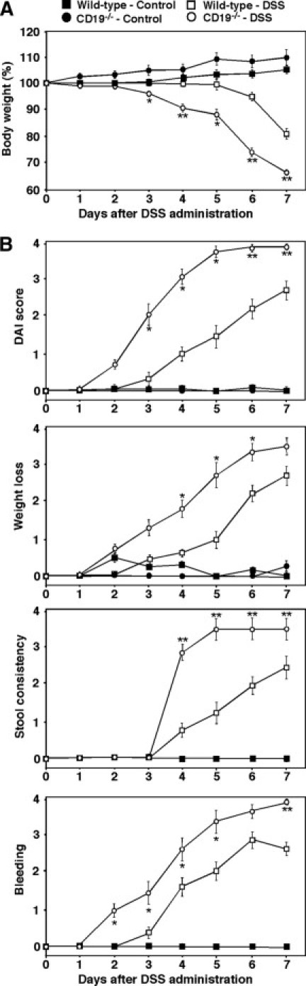
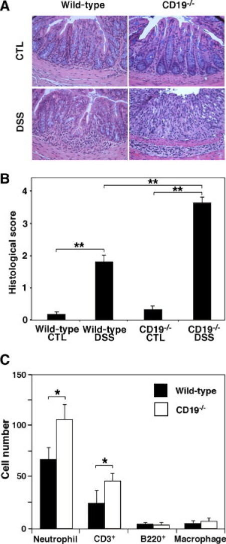
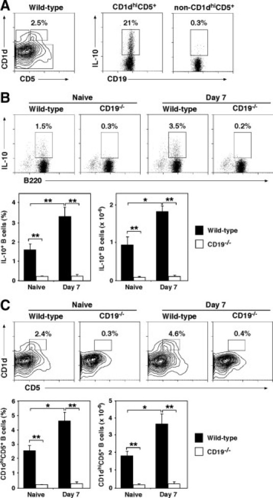
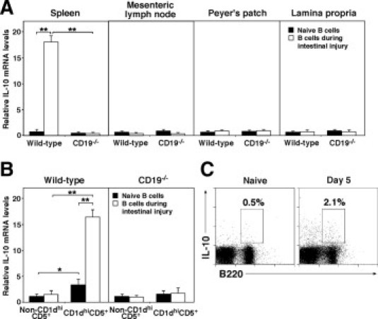
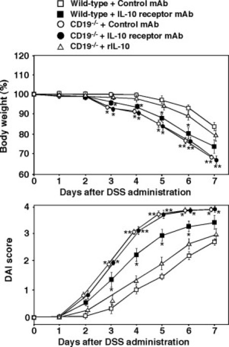
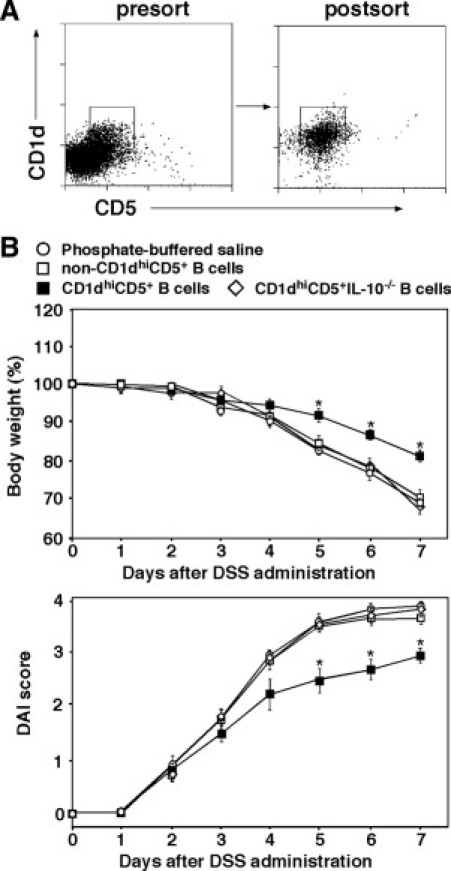
B cells mediate multiple functions that influence immune and inflammatory responses. In mice, the addition of dextran sulfate sodium (DSS) to drinking water leads to immediate intestinal injury. Dextran sulfate sodium-induced intestinal injury serves as an experimental animal model for human ulcerative colitis. The contribution of B cells to DSS-induced intestinal injury is unclear. In this study, we show that DSS-induced intestinal injury was more severe in CD19-deficient (CD19(-/-)) mice than in wild-type mice. These inflammatory responses were negatively regulated by a unique IL-10-producing CD1d(hi)CD5(+) regulatory B cell subset (B10 cells) that was absent in CD19(-/-) mice and represented only 1% to 2% of splenic B220(+) cells in wild-type mice. Remarkably, adoptive transfer of these B10 cells from wild-type mice reduced inflammation in CD19(-/-) mice in an IL-10-dependent manner. These results demonstrate that IL-10 production from regulatory B10 cells regulates DSS-induced intestinal injury. These findings may provide new insights and therapeutic approaches for treating ulcerative colitis.


Figures

Figure 1
Figure 1
Increased severity of DSS-induced intestinal injury in CD19−/− mice. Wild-type and CD19−/− mice ingested either DSS solution or normal drinking water (control). The severity of intestinal injury was evaluated by quantitatively measuring body weight (A) and DAI scores (B). The DAI scores were based on weight loss, stool consistency, and bleeding. Values represent the mean ± SEM from four or more mice per group. Significant differences between sample means are indicated: *P < 0.05 and **P < 0.01. Similar results were obtained in at least two independent experiments.
Figure 2
Figure 2
CD19 deficiency enhanced the severity of DSS-induced intestinal injury. Colon sections were harvested from wild-type and CD19−/− mice after ingestion of either DSS solution or normal drinking water for 7 days; sections were stained with H&E. A: Representative colon sections from wild-type and CD19−/− mice at 7 days after induction of intestinal injury. B: Histological sections were blindly scored on a scale of 0 to 4 for severity of colitis. C: The numbers of neutrophils, CD3+ T cells, B220+ B cells, and F4/80+ macrophages per one field of view (×400) were counted. Values represent the mean ± SEM from four or more mice per group. B and C: Significant differences between sample means are indicated: *P < 0.05 and **P < 0.01. Results represent one of two independent experiments producing similar results. CTL, control.
Figure 3
Figure 3
IL-10 production by splenic B cells correlates with suppression of DSS-induced intestinal injury. A: CD1dhiCD5+ B cells are the predominant IL-10–producing B-cell subset. Splenocytes from wild-type mice were cultured with lipopolysaccharide (LPS), phorbol 12-myristate 13-acetate (PMA), ionomycin, and monensin for 5 hours before permeabilization and staining using CD19, CD1d, CD5, and IL-10 mAbs. IL-10 production by CD19+ B cells within the CD1dhiCD5+ and non–CD1dhiCD5+ subpopulations is shown, with the proportions of IL-10+ cells within the indicated gates shown. B: Splenic IL-10–producing B-cell proportions and absolute numbers increase during DSS-induced intestinal injury in wild-type mice but not in CD19−/− mice. Splenocytes were isolated from wild-type and CD19−/− mice before and 7 days after the induction of intestinal injury. B220+ splenocytes were purified from wild-type and CD19−/− mice. Purified B cells were incubated in the presence of lipopolysaccharide, phorbol 12-mynstate 13-acetate, ionomycin, and monensin for 5 hours. B cells were stained with B220 mAb. After permeabilization, the cells were stained with IL-10 mAb. Representative results demonstrate the proportion of IL-10–producing cells among the total B220+ B cells within the indicated gates. Bar graphs indicate the mean ± SEM percentages and numbers of B cells that produce IL-10. C: CD1dhiCD5+ B-cell proportions and absolute numbers increase during DSS-induced intestinal injury in wild-type mice but not in CD19−/− mice. Splenocytes were isolated from wild-type and CD19−/− mice before and 7 days after the induction of intestinal injury and analyzed for CD1d, CD5, and B220 expression by immunofluorescence staining with flow cytometry analysis. Representative results demonstrate the proportion of CD1dhiCD5+ B cells among the total B220+ B cells within the indicated gates. Bar graphs indicate the mean ± SEM percentages and numbers of CD1dhiCD5+ B cells. B and C: Significant differences between sample means are indicated: *P < 0.05 and **P < 0.01. All results represent two or more independent experiments, with four mice in each group.
Figure 4
Figure 4
A: IL-10 production by wild-type and CD19−/− mice during DSS-induced intestinal injury. B220+ cells were purified from the spleen, mesenteric lymph node, Peyer's patches, and intestinal lamina propria of DSS-treated and control mice. B: Splenic CD1dhiCD5+ or non–CD1dhiCD5+ B cells were purified from naïve or DSS-treated mice by cell sorting. C: IL-10 production by circulating B cells from wild-type mice during DSS-induced intestinal injury. Blood mononuclear cells from three mice were pooled and cultured with lipopolysaccharide, phorbol 12-myristate 13-acetate, ionomycin, and monensin for 5 hours before permeabilization and staining using B220, CD1d, CD5, and IL-10 mAbs. Values represent the proportion of IL-10–producing cells among the total B cells. A and B: Transcript levels were quantified by real-time PCR analysis and were normalized to the internal control. Significant differences between sample means are indicated: *P < 0.05 and **P < 0.01. All results represent two or more independent experiments, with four mice in each group.
Figure 5
Figure 5
The suppression of DSS-induced intestinal injury is IL-10 dependent. The DSS-induced intestinal injury in wild-type or CD19−/− mice treated with control or IL-10 receptor–specific mAb on days 0 and 3. A group of CD19−/− mice was treated with rIL-10 on days 0, 2, 4, and 6. Significant differences between sample means are indicated: *P < 0.05 and **P < 0.01. All results represent two or more independent experiments, with four mice in each group.
Figure 6
Figure 6
Regulatory CD1dhiCD5+ B10 cells suppress disease symptoms in DSS-induced intestinal injury. A: Representative results showing splenic B220+ cells from wild-type mice sorted into the CD1dhiCD5+ B-cell subset. B: CD1dhiCD5+ or non–CD1dhiCD5+ B cells were purified from naïve wild-type mice by cell sorting. Purified cells were transferred into CD19−/− mice. The DSS was administered to the recipient mice 48 hours after transfer. Significant differences between PBS-treated mice versus other groups are indicated: *P < 0.05. Values represent the mean ± SEM from four or more mice in each group.

Similar articles

Cited by

References

    1. Fiocchi C. Inflammatory bowel disease: etiology and pathogenesis. Gastroenterology. 1998;115:182–205. - PubMed
    1. Hibi T., Ogata H. Novel pathophysiological concepts of inflammatory bowel disease. J Gastroenterol. 2006;41:10–16. - PubMed
    1. Sands B.E. Inflammatory bowel disease: past, present, and future. J Gastroenterol. 2007;42:16–25. - PMC - PubMed
    1. Okayasu I., Hatakeyama S., Yamada M., Ohkusa T., Inagaki Y., Nakaya R. A novel method in the induction of reliable experimental acute and chronic ulcerative colitis in mice. Gastroenterology. 1990;98:694–702. - PubMed
    1. Cooper H.S., Murthy S.N., Shah R.S., Sedergran D.J. Clinicopathologic study of dextran sulfate sodium experimental murine colitis. Lab Invest. 1993;69:238–249. - PubMed

Publication types