. 2011 Feb;178(2):754-63.
doi: 10.1016/j.ajpath.2010.10.015.

Tenascin C induces epithelial-mesenchymal transition-like change accompanied by SRC activation and focal adhesion kinase phosphorylation in human breast cancer cells

Affiliations

Tenascin C induces epithelial-mesenchymal transition-like change accompanied by SRC activation and focal adhesion kinase phosphorylation in human breast cancer cells

Keiki Nagaharu et al. Am J Pathol. 2011 Feb.

Abstract

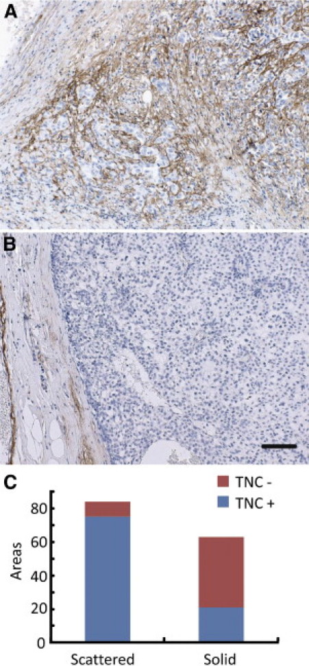
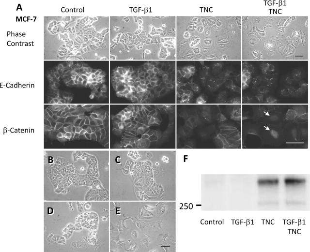
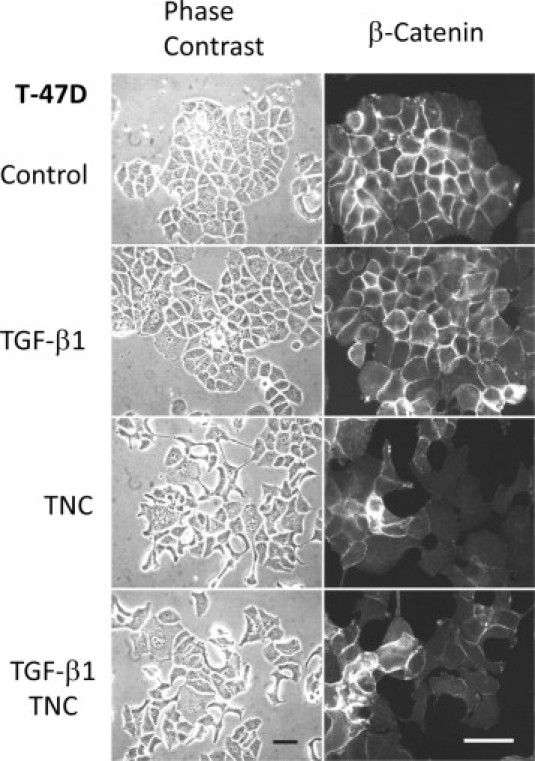
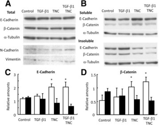
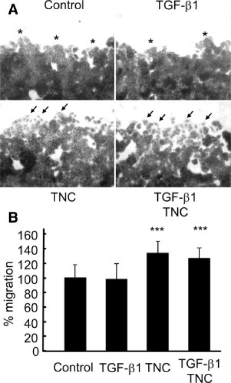
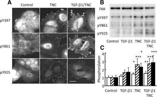
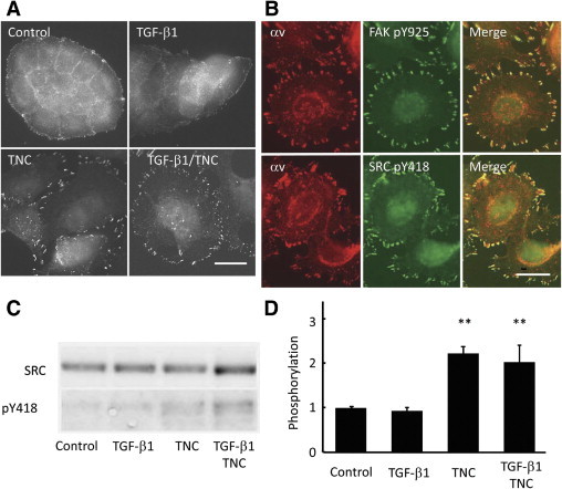
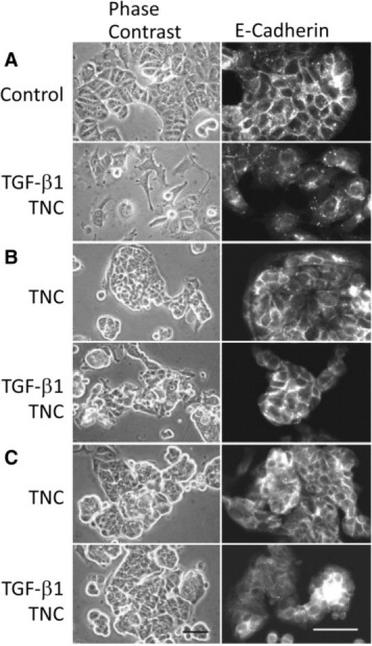
Tenascin C (TNC) is an extracellular matrix glycoprotein up-regulated in solid tumors. Higher TNC expression is shown in invading fronts of breast cancer, which correlates with poorer patient outcome. We examined whether TNC induces epithelial-mesenchymal transition (EMT) in breast cancer. Immunohistochemical analysis of invasive ductal carcinomas showed that TNC deposition was frequent in stroma with scattered cancer cells in peripheral margins of tumors. The addition of TNC to the medium of the MCF-7 breast cancer cells caused EMT-like change and delocalization of E-cadherin and β-catenin from cell-cell contact. Although amounts of E-cadherin and β-catenin were not changed after EMT in total lysates, they were increased in the Triton X-100-soluble fractions, indicating movement from the membrane into the cytosol. In wound healing assay, cells were scattered from wound edges and showed faster migration after TNC treatment. The EMT phenotype was correlated with SRC activation through phosphorylation at Y418 and phosphorylation of focal adhesion kinase (FAK) at Y861 and Y925 of SRC substrate sites. These phosphorylated proteins colocalized with αv integrin-positive adhesion plaques. A neutralizing antibody against αv or a SRC kinase inhibitor blocked EMT. TNC could induce EMT-like change showing loss of intercellular adhesion and enhanced migration in breast cancer cells, associated with FAK phosphorylation by SRC; this may be responsible for the observed promotion of TNC in breast cancer invasion.


Figures

Figure 1
Figure 1
Dense TNC deposition is associated with an invading phenotype of human breast cancer cells. A: Invading cancer cells form small clusters scattered in TNC-positive cancer stroma. B: With large clusters showing smooth peripheral margins, TNC is not deposited in the stroma. Scale bar = 50 μm. C: TNC staining was frequently positive in the stroma with scattered cancer cells. The difference in positivity of TNC staining between solid and scattered types was significant (P < 0.0001).
Figure 2
Figure 2
TNC induces EMT-like change and E-cadherin/β-catenin translocation in MCF-7 cells. A: In the control and TGF-β1 treatment cases, MCF-7 cells form large clusters with tight intercellular connections. TNC was added to the medium when the cells were plated. Treatment for 64 hours induced cell-cell dissociation, and combined treatment with TGF-β1 and TNC resulted in stronger morphological change (top). On immunofluorescence of control cells, E-cadherin (middle) and β-catenin (bottom) are localized at plasma membranes of cell-cell junctions. TGF-β1 treatment did not affect localization, but TNC treatment reduced the membranous localization and the combined treatment–induced β-catenin translocation into nuclei (arrows). Scale bars = 50 μm. B: EMT change is reversible because the morphology was restored to the normal epithelial phenotype within 3 days after replacement of the normal medium. C–D: As a negative control, addition of a supernatant of TNC solution after immunoprecipitation by TNC antibody without TGF-β1 (C) or with TGF-β1 (D) did not cause phenotypic change. E: Addition of a neutralizing antibody to TGF-β (1D11, 50 μg/ml) into the medium containing TNC alone was examined, but this did not impede the EMT, indicating a direct effect of TNC on EMT. Scale bar = 50 μm. F: Immunoblot analysis of the lysate showed TNC deposition on cell surfaces and substrata in TNC- and TGF-β1/TNC-treated groups, although TNC was barely detectable in control and TGF-β1 alone groups.
Figure 3
Figure 3
TNC also induces EMT-like change in T-47D cells. Breast cancer cell line T-47D showed EMT-like change by TNC and TGF-β1/TNC treatments for 5 to 6 days. The EMT was exhibited at the margin of cell cluster, to a lesser extent than for MCF-7 cells (left). Immunofluorescence exhibited reduced β-catenin staining of intercellular contacts in the cells moving outside the cluster (right). Scale bars = 50 μm.
Figure 4
Figure 4
Immunoblot results showing translocation of E-cadherin and β-catenin to cytoplasm in MCF-7 cells after TNC treatment. A: Total amounts of E-cadherin and β-catenin extracted with SDS buffer do not differ among the samples. Expression of mesenchymal markers N-cadherin and vimentin is not up-regulated. B–D: On Triton X-100 fractionation [0.5% Triton X-100, 2.5 mmol/L EGTA, 5 mmol/L MgCl2, and 50 mmol/L PIPES (pH 6.2) at 37°C], the soluble fractions (open columns) show cytoplasmic/cytosolic localization, whereas the insoluble fractions (filled columns) demonstrate binding to the adherens junctions held by preserved cytoskeleton. TNC and TGF-β1/TNC treatments increased cytoplasmic/cytosolic fractions of E-cadherin (C) and β-catenin (D), indicating dissociation from the cytoskeleton/membrane. *P < 0.05.
Figure 5
Figure 5
TNC induces scattered migration of MCF-7 cells from the edges and promotes migration in wound healing assay. A: TNC and TGF-β1/TNC treatments showed an increase in the number of scattered migrating cells (arrows) from the wound edges, whereas cells in the control and after TGF-β1 treatment groups exhibited tight clustering (asterisks). Scale bar = 50 μm. B: Migration of cells with TNC and TGF-β1/TNC treatments was significantly faster than that in the control or TGF-β1 treatment groups. Area newly covered by the migrating cells after wounding in the control group is denoted as 100%. Thirty-six fields per condition were examined. ***P < 0.001.
Figure 6
Figure 6
TNC induces FAK phosphorylation. A: Immunofluorescent staining of MCF-7 cells with antibodies against phosphorylated FAK demonstrates an increase in the number of adhesion plaques positive for pY397, pY861, and pY925 with TNC and combined TGF-β1/TNC treatments. Bright fluorescence for FAK pY397 was visible in ruffled membranes and observed more frequently in the TNC- and TGF-β1/TNC-treated cells (asterisks). Scale bar = 20 μm. B: Immunoblot analyses of MCF-7 lysates using antibodies against FAK and the phosphorylated sites also show enhanced phosphorylation of FAK pY397, pY861, and pY925 after TNC and the combined treatments. C: Quantified data also showed that phosphorylation of FAK Y397 (open column), Y861 (striped columns), and Y925 (filled columns) in TNC-treated groups is significantly increased. *P < 0.05, **P < 0.01, ***P < 0.001.
Figure 7
Figure 7
TNC induces SRC activation accompanied by phosphorylation at Y418. A: Immunofluorescent staining of MCF-7 cells with antibodies against SRC pY418 demonstrates an increase in the number of adhesion plaques positive for pY418 with TNC and combined TGF-β1/TNC treatments. Scale bar = 20 μm. B: Immunofluorescent staining of FAK pY925 or SRC pY418 colocalizes with αv-positive adhesion plaques. Scale bar = 20 μm. C and D: Immunoblot analyses using antibodies against phosphorylated SRC show enhanced phosphorylation of Y418 after TNC and the combined treatments. **P < 0.01.
Figure 8
Figure 8
A neutralizing antibody against αv integrin and an SRC kinase inhibitor inhibit EMT change after the TNC alone and TGF-β1/TNC treatments. A: Phase contrast microscopy (left) and E-cadherin immunofluorescence (right) of MCF-7 cells without any treatment and after TGF-β1/TNC treatment are exhibited as controls. B: A neutralizing antibody against αv integrin (AV1) blocks EMT change after TNC and TGF-β1/TNC treatments. The attached cells on the substratum are reduced in number by AV1 treatment. Immunofluorescence staining of E-cadherin was preserved in cell-cell contacts. C: The SRC kinase inhibitor (pp2) also inhibits the phenotypic change, with a tendency to form rounded clusters. Scale bars = 50 μm.

References

    1. Savagner P. Leaving the neighborhood: molecular mechanisms involved during epithelial-mesenchymal transition. Bioessays. 2001;23:912–923. - PubMed
    1. Thiery J.P., Acloque H., Huang R.Y., Nieto M.A. Epithelial-mesenchymal transitions in development and disease. Cell. 2009;139:871–890. - PubMed
    1. Baum B., Settleman J., Quinlan M.P. Transitions between epithelial and mesenchymal states in development and disease. Semin Cell Dev Biol. 2008;19:294–308. - PubMed
    1. Yang J., Weinberg R.A. Epithelial-mesenchymal transition: at the crossroads of development and tumor metastasis. Dev Cell. 2008;14:818–829. - PubMed
    1. Thompson E.W., Newgreen D.F., Tarin D. Carcinoma invasion and metastasis: a role for epithelial-mesenchymal transition? Cancer Res. 2005;65:5991–5995. - PubMed

Publication types

MeSH terms